
Case contribution: Dr Radhiana Hassan
Clinical:
- A 49 years old lady
- Known case of left breast cancer, mastectomy done 5 years ago
- Completed radiotherapy and chemotherapy
- Presented with right periorbital swelling and diplopia
- No history of trauma, no fever.
- No constitutional symptoms
CT scan findings:
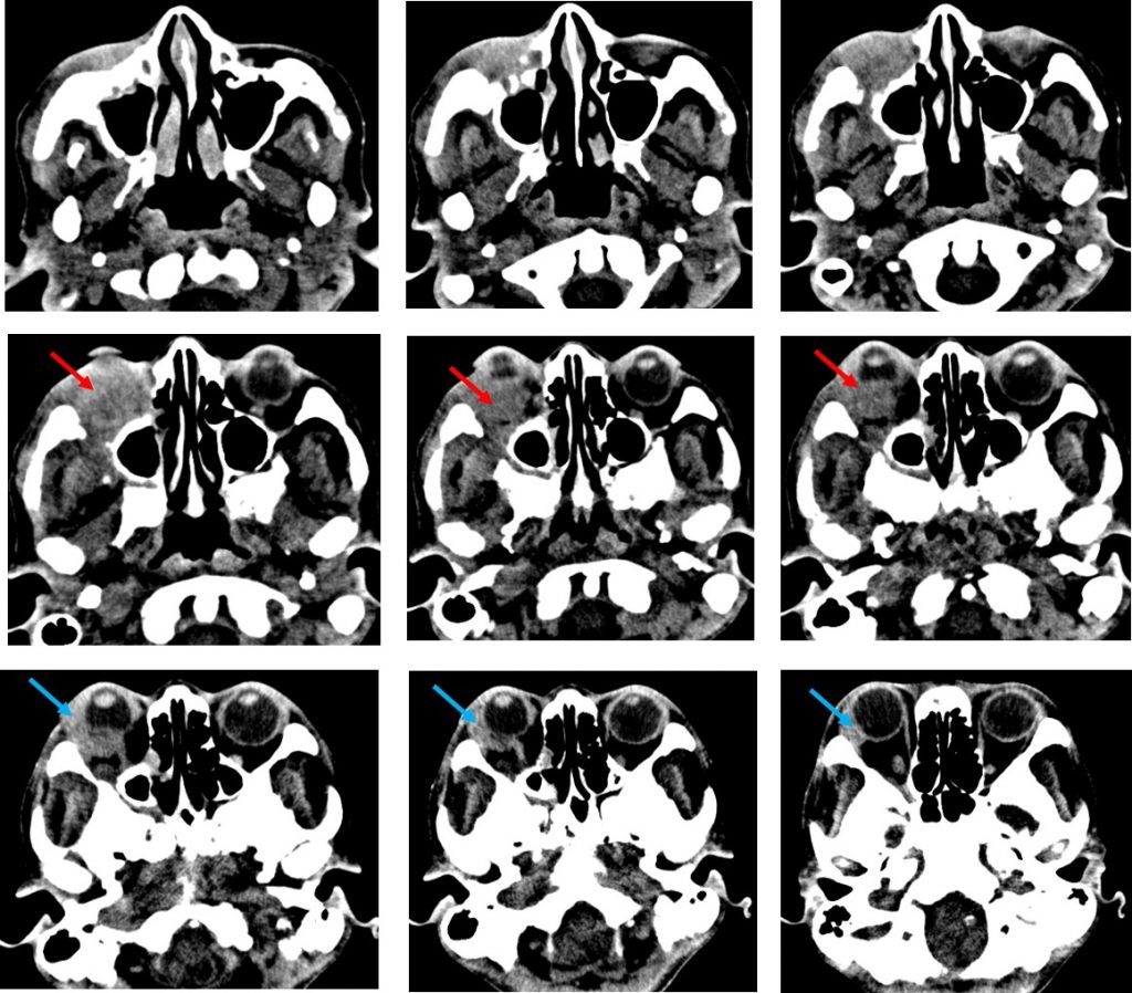
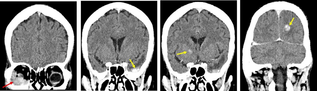
- Mildly enhancing soft tissue mass in the extraconal region of right orbital cavity (red arrows). It is located at lateral and inferior quadrant. No calcification seen.
- The right lacrimal gland also looks bulky (blue arrows).
- Extraocular muscles are normal in size and symmetrical in appearance.
- Optic nerve is normal.
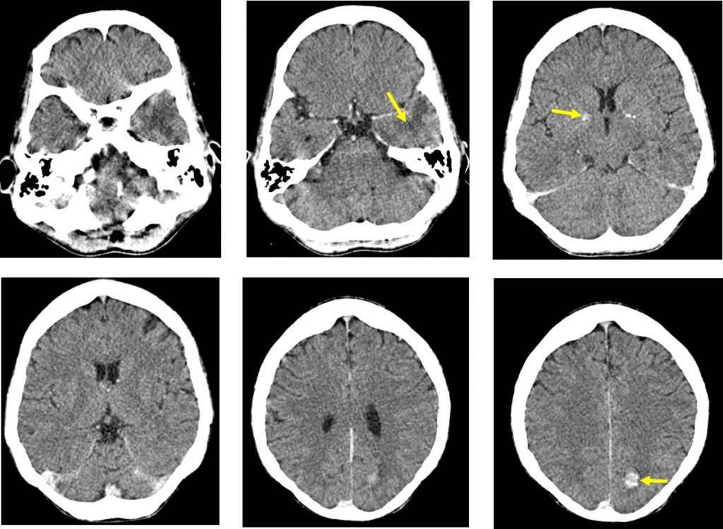
- In the brain, there are a few enhancing lesions in the left temporal lobe and left occipital lobe (yellow arrows).
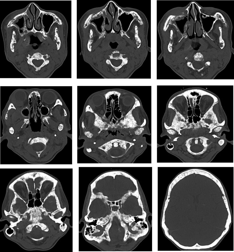
- There is extensive lytic sclerotic lesions of the skull vault and facial bones.
Progress of patient:
- Subsequent CT thorax shows multiple lung metastasis.
- Also had multiple liver metastasis.
Diagnosis: Multiple metastasis from breast cancer.
Discussion:
- Orbital metastases are infrequent; about 6% of all orbital mass biopsied are metastatic in origin.
- Extraocular metastases accounts for 2-11% of all orbital neoplasms.
- Breast carcinoma and lung carcinoma are the most common primary tumours of orbital metastasis.
- Not infrequently, patients have silent brain lesion when they present with orbital disease. Thus, imaging of whole brain should be included.
- Extraocular metastasis are usually unilateral and only infrequently primarily involve the extra-ocular muscles.
- The superior and lateral extraconal quadrant is the most frequently involved although any parts can be involved.
Recent Comments