Case contribution: Dr Radhiana Hassan
Clinical:
- A 21 years old man, history of cyanotic heart disease and operation done 5 years ago. Post op NYHA I.
- Involved in MVA ( motorcycle versus a car)
- Complaint of left lumbar pain and hematuria
- Bedside ultrasound shows left renal injury
- CT for further assessment


CT scan findings:
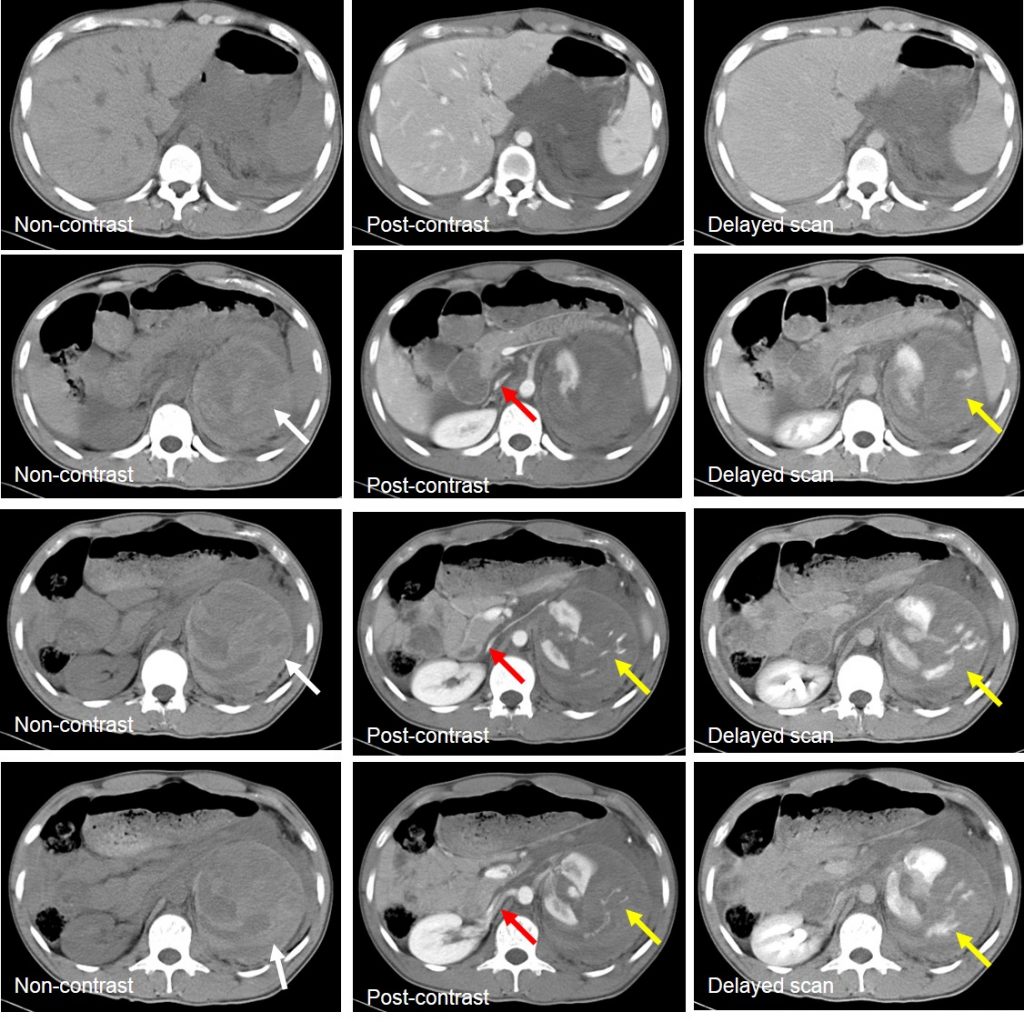
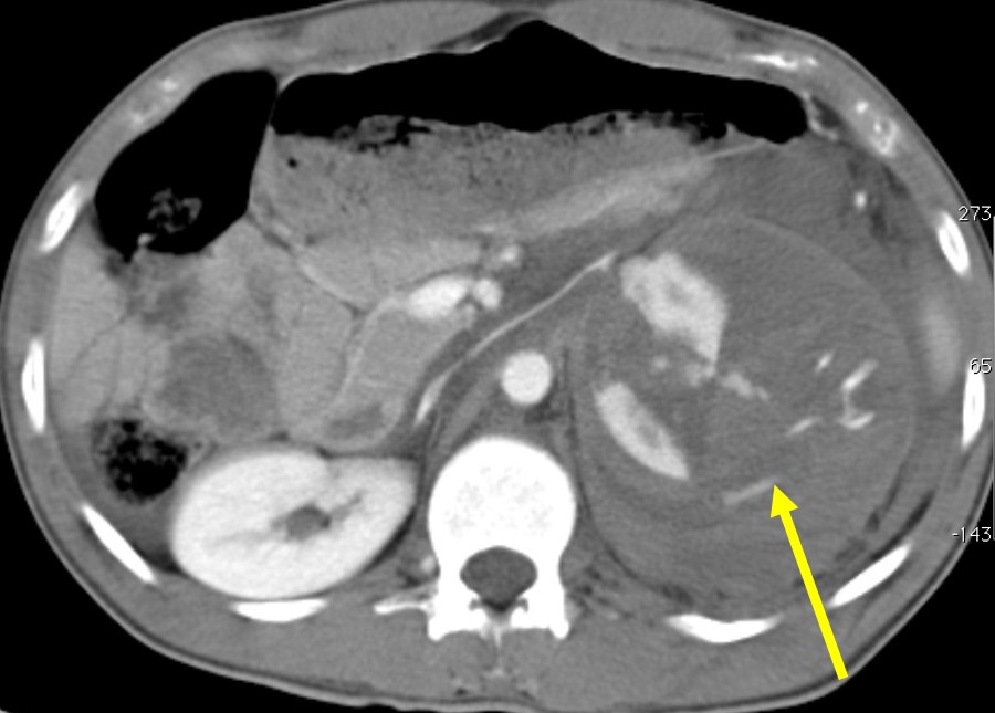
- Multiple deep lacerations of left renal cortex extending to collecting system centrally
- There is associated large perinephric hematoma (white arrows)
- It causes displacement of splenic artery and veins, pancreas and left renal vein anteriorly
- Left renal perfusion and excretion is preserved
- There is contrast extravasation suggestive of active hemorrhage (yellow arrows)
- Flat IVC (red arrows) suggestive of hypovolemia state
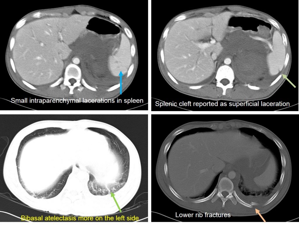
- Small splenic intraparenchymal lacerations seen
- Fractures of lower ribs on the left side
Intraoperative findings:
- Lacerations at lateral aspect of left kidney, extending to hilum,
- Active hemorrhage with extensive hemoperitoneum. Blood loss 2200ml.
- Spleen normal. Splenic cleft seen at lateral surface
- Left nephrectomy performed.
Diagnosis: Shattered left kidney with active hemorrhage
Progress of patient:
- Active CPR done during surgery for 3 minutes
- A total of 8pint PC, I cycle DIVC regime was transfused.
- Patient was discharged well 2 weeks later
- Discharged, Hb=11.9 and review after 2 weeks show patient was well
Discussion:
- Renal injuries count for about 10% of abdominal trauma
- Computed tomography (CT) is the modality of choice in the evaluation of blunt renal injury
- CT can provide precise delineation of a renal laceration, help determine the presence and location of a renal hematoma
- It can demonstrate injury with or without active arterial extravasation,
- It can also show the presence of urinary extravasation or of devascularized segments of renal parenchyma.
- Most important, CT can help differentiate trivial injuries from those requiring intervention.
- Shattered kidney is considered Grade V in AAST grading scale
