
Case contribution: Dr Radhiana Hassan
Clinical:
- A 4 years old girl
- Presented with vomiting after a fall
- Urgent CT scan shows a mass lesion at sellar region
- Referred for further investigation and management
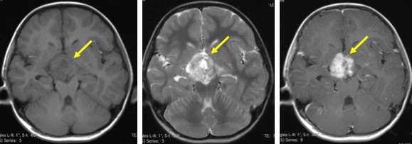
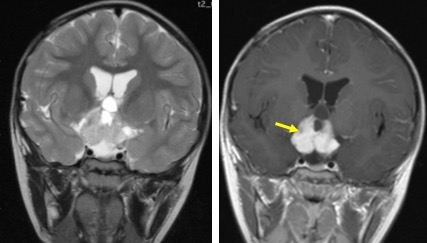
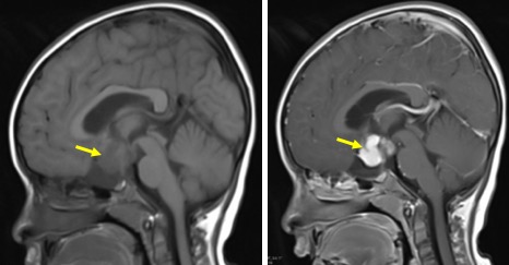
MRI findings:
- A well-defined mass lesion at suprasellar region (yellow arrows)
- It shows mixed signal intensity and predominantly solid
- Vivid enhancement of solid component is seen
- No intralesional calcification or hemorrhage
- Sella and pituitary is normal
- No abnormal meningeal/dural enhancement
Diagnosis: Pilocytic astrocytoma (HPE proven)
Discussion:
- Pilocytic astrocytoma at sella/suprasella region is known as optic pathway glioma
- It typically present in children, accounting for 10-15% of supratentorial tumors in this age group
- Males and females are approximately equally affected
- In adults, optic nerve gliomas do occur but are very rare and usually aggressive tumors
- Association with NF 1 in 10-63%
Recent Comments