Case contribution: Dr Radhiana Hassan
Clinical:
- A 60 years old lady
- No known medical illness
- Chest radiograph performed for preoperative assessment of another condition

X-ray findings:
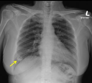
- There is a lobulated calcified lesion seen at right lower zone (yellow arrow). Central lucency is seen within this lesion.
- The right lower zone of the lung around the lesion appears unremarkable.
- No pleural effusion. No evidence of adjacent pleural thickening is seen.
- The adjacent ribs are also preserved.
- The rest of the visualised lung appears normal.
- No hilar or mediastinal mass.


Mammogram findings:
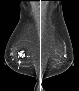
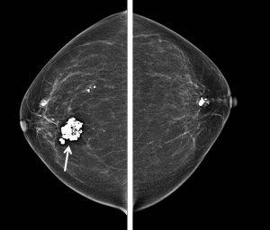
- Both breasts are almost entirely fatty (BIRADS composition A).
- There are few lesions with coarse calcifications are seen in the right breast.
- The largest is seen at upper mid-quadrant, with a popcorn-like calcifications.
- Similarly in the left breast, there is a low density lesion with coarse calcification seen at upper mid-quadrant.
- No suspicious cluster of microcalcifications.
- No skin thickening or nipple retraction.
- Small right axillary node is seen

Ultrasound findings:
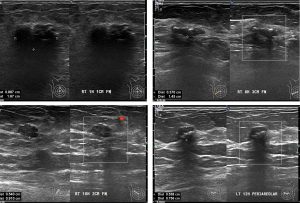
- There are multiple lesions in both breasts
- Some shows intraleisonal calcificaiton as noted on mammogram
- Some are without calcificaitons
- No suspicious feature of these lesions
- No significant axillary lymphadenopathy.
Diagnosis: Benign lesions in both breasts -involuting fibroadenomas (presumptive diagnosis).
Discussion: Popcorn calcification
- Calcifications that are benign at mammography are typically larger, coarser, round with smooth margins and more easily seen than malignant calcifications.
- Popcorn calcifications are classic, large, >2-3 mm in greatest dimension
- It is produced by involuting fibroadenomas
- It can be seen in stages, initially as peripheral calcification, later it appears confluent and later become almost completely calcified.
