
Case contribution: Dr Radhiana Hassan
Clinical:
- A 58 years old
- History of right breast cancer 9 years ago
- WLE, axillary clearance and radiotherapy treatment (in another hospital)
- No chemotherapy or hormonal therapy after that
- Complaint of recently felt left breast lump
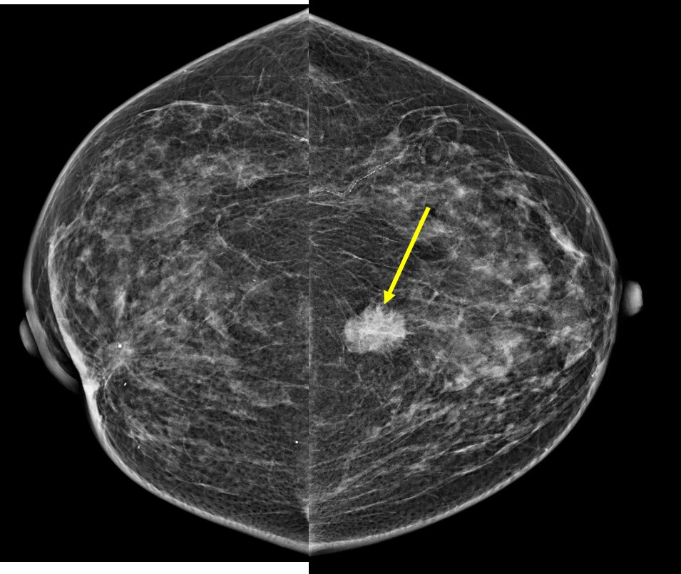
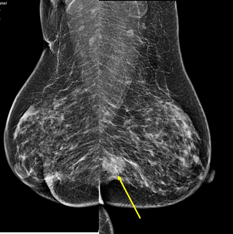
Mammogram findings:
- Moderately dense breasts (BIRADS B) with symmetrical pattern
- A high density mass at left lower inner quadrant (arrows)
- Lobulated appearance with no associated suspicious clustered microcalcification
- A density and stromal distortion at right breast from previous operation
- No skin thickening. No abnormal axillary nodes.
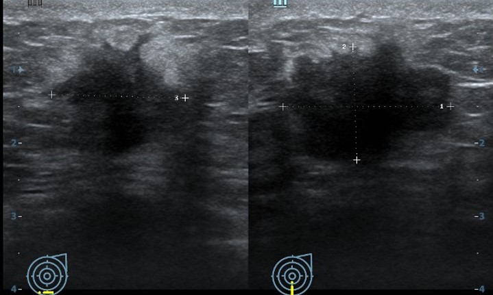
Ultrasound findings:
- A suspicious mass at Lt6H measuring 23x18x16 mm
- It shows irregular outline with finger-like infiltration
- Posterior shadowing is also seen
- No abnormal vascularity
- No abnormal axillary node
HPE: invasive carcinoma, no special type
Diagnosis: Metachronous breast cancer
Discussion:
- Metachronous breast cancers are two breast cancers that occur in either breast in two different time periods.
- Others defined metachronous as those diagnosed after 6 months from the first BC diagnosis in the contralateral breast or in the same breast but with different histology.
- The prevalence of metachronous breast cancer is 5-7%
- Bilaterality is greatest with invasive lobular carcinoma
- Metastasis to the breast from opposite breast is one of the cause especially with other evidence of metastasis
- The risk of developing metachronous contralateral carcinoma can be 0.9% per year, with a cumulative risk of 12% at 15 years. Thus, support continuous long term cancer surveillance.
Recent Comments