Case contribution: Dr Radhiana Hassan
Clinical:
- A 49-year-old female, underlying perforated sigmoid colon cancer.
- CT scan done for staging.
- Incidental finding of vertebral lesion


CT scan findings:
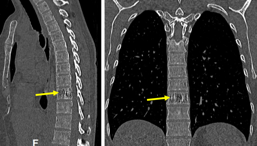
- An ill-defined lytic changes seen at T10 vertebrae
- There are striated vertical densities within it (yellow arrows)
- On axial images the vertical densities are seen as rounded foci giving rise to ‘polka dot’ sign
- No cortical break. No surrounding soft tissue mass
Diagnosis: Vertebral hemangioma (jail bar and polka dot signs)
Discussion:
- The jail bar sign refers to the vertically striated appearance seen in vertebrae due to thickening of the bony trabeculae.
- It is named as the appearance mimics the appearance of prison jail bars.
- This appearance is strongly associated with vertebral hemangiomas.
- It is also known as ‘colduroy cloth’ sign.
- Axial CT will show a ‘polka-dot’ sign due to the thickened vertebral trabeculae. On CT the dots are white on a black fatty background.
- On MRI they are black dots on a white background (on non-fat-suppressed T1 or T2-weighted images).
- ‘Polka dot’ sign is also known as ‘salt and pepper’ sign for obvious reasons.
