Image:

Questions:
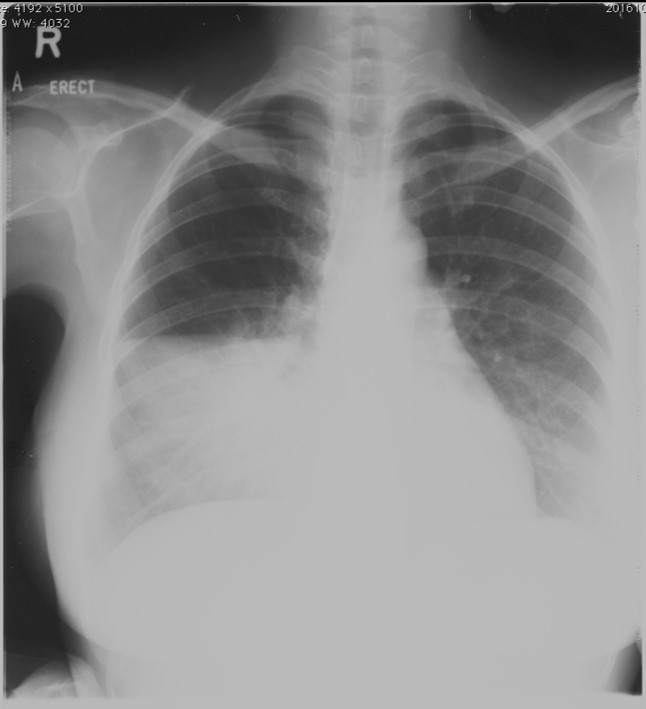
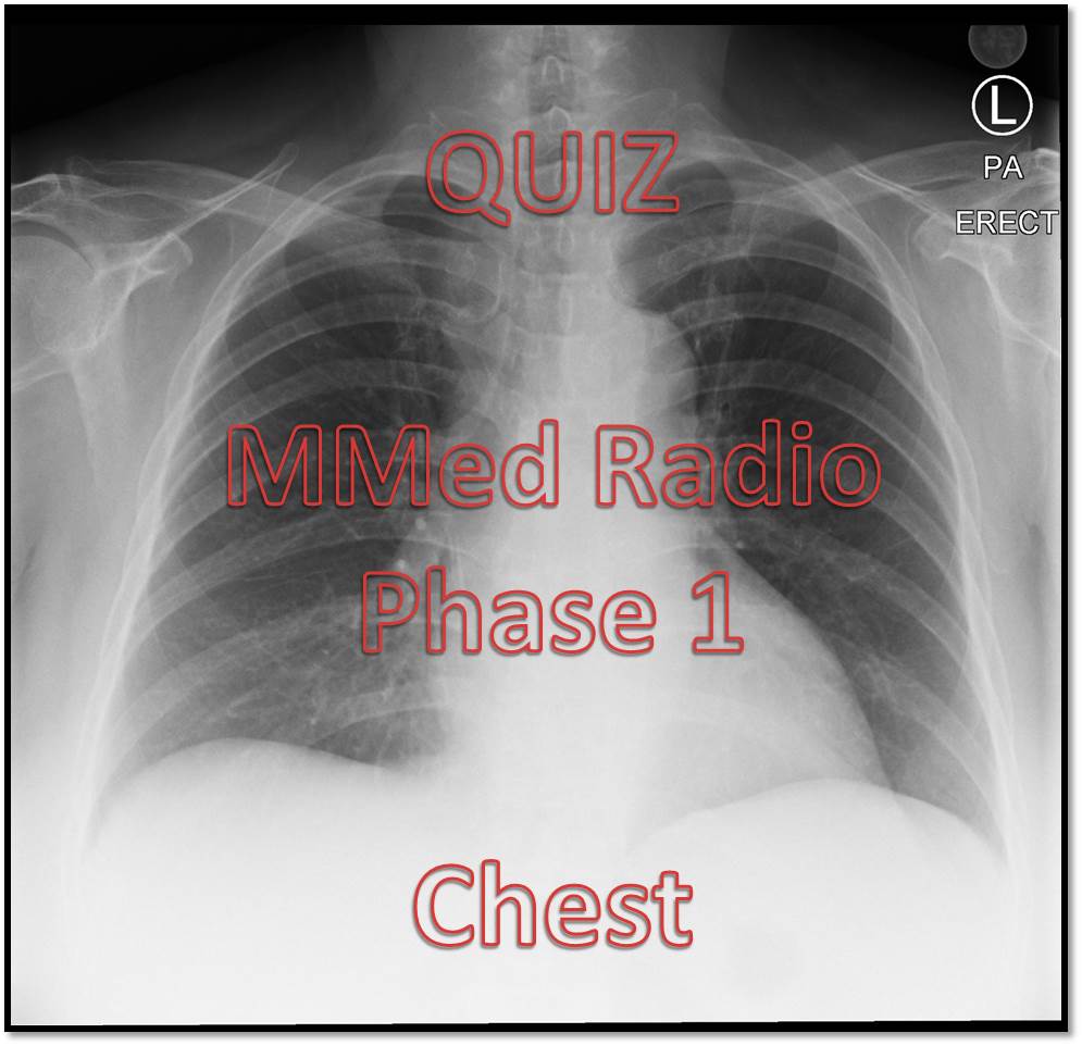
- Name the examination.
- Based on the image, which lung lobe is affected in this case?
- Give reasons for your answer in Question 2.
- How to confirm the location of the affected lobe?
Answers:
- Chest radiograph, PA erect view.
- Right middle lobe.
- The affected region has clear demarcation superiorly which is bounded by horizontal fissure and right cardiac margin is obliterated (sillhoutte sign).
- Lateral chest radiograph.
