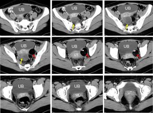
Clinical:
- A 24 years old lady
- Presented with abdominal discomfort
- Normal menstrual cycle

CT scan findings:
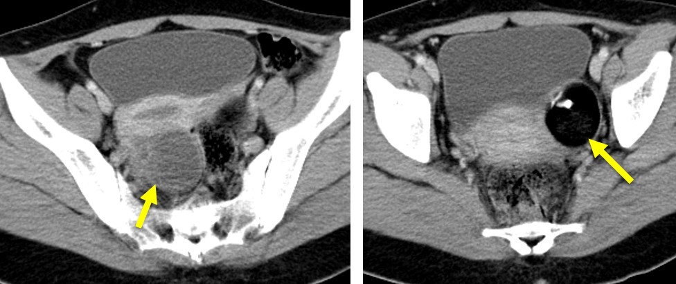
- There is a well defined rounded lesion at left adnexal area (red arrows). The lesion is predominantly fat (-100HU). A small calcified mural nodule is seen within the lesion. There is no enhancement post contrast
- Another lesion which is smaller and mainly cystic seen on the right side (yellow arrows). A few thin septae within which shows slight contrast enhancement.
- The fat plane with surrounding structures are still preserved.
- Uterus is normal.
Intra-operative findings:
- Left ovarian cyst measuring 6×4 cm. Punctured one hole, secrete sebum-like fluid, removed with intact capsule. Ovarian reconstruction done.
- A 4 cm right ovarian endometrioma seen. Ruptured during manipulation, released chocolate material. Stuck at POD. Cyst wall removed. Ovarian reconstruction done.
- Minimal peritoneal fluid. No endometriotic spot
HPE findings:
- Macroscopy: specimen labelled as left ovarian cyst consists of intact cystic mass measuring 60x43x40 mm. Cut section shows a uniloculated cyst filled with yellowish cheesy white material and clumps of hair. The cyst wall measure 1 mm in thickness. There is hard structure within the lesion, probably a tooth. Another specimen labelled as right cyst wall consists of multiple pieces of greyish tissue measuring 35 mm.
- Microscopy: Sections of left ovarian cyst shows cyst wall are lined by keratinized stratified squamous epithelium. The underlying stroma show presence of skin adnexa and skeletal muscle bundles. No immature component or malignancy is seen. Sections of right adnexal cyst shows multiple cyst walls lined by luteinized cells. Focal area of haemorrhage is noted. No evidence of malignancy.
- Interpretation: Left ovarian cystic mature teratoma and right corpus luteal cyst.
Diagnosis: Mature ovarian teratoma and corpus luteal cyst.
