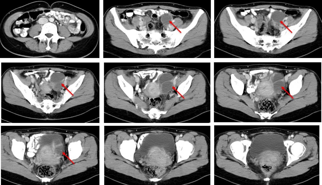
Case contribution: Dr Radhiana Hassan
Clinical:
- A 41 years old
- Presented with acute onset abdominal pain
- No fever, no bowel habit change.
- No constitutional symptoms

CT scan findings:
- The uterus is bulky iwth inhomogenous parenchymal enhancement
- There is a complex cystic-solid lesion in the left adnexa
- The lesion measures 6x5x3 cm.
- There is enhancing region at the superomedial surface.
- Parametrial streakiness with few shotty nodes
- No ascites
Intra-operative findings:
- Left endometrioma about 6×6 cm
- POD is obliterated
- Adhesion of the uterus to the rectum posteriorly
- The left fallopian tube and left ovary embedded to the posterior of uterus
- Right ovary and right fallopian tube is normal
- Endometriotic spots noted at the fundus of uterus.
- No endometriotic spots at the bowel
HPE findings:
- Macroscopy: specimen labelled as left ovary and cyst wall consist of ruptured cystic lesion measuring 38x25x25 mm and 2-5 mm thickness.
- Microscopy: section shows multiple pieces of ovarian cyst wall with areas of hemorrhage. The cyst walls are lined by hemorrhagic endometrial stroma with some areas show presence of endometrial epithelium. A few foci of microcalcification are noted. No evidence of malignancy.
- Interpretation: compatible with endometriotic cysts
Diagnosis: Endometriotic cysts
Discussion:
- Endometriotics cysts are noncancerous, fluid-filled cysts that typically form deep within the ovaries.
- It is also known as chocholate cyst due to its appearance of brown, tar-like appearance, looking something like melted chocolate. The colour comes from old menstrual blood and tissue that fills the cavity of the cyst.
- An endometriotic cyst can affect one or both ovaries, and may occur in multiples or singularly.
- It occurs in 20-40% of women having endometriosis.
- Endometriotic cysts can place a a woman of reproductive age at a higher risk for ovarian cancer, can cause pelvic pain, contribute to infertility, decrease ovarian function and interfere with assisted reproductive technologies.
