Clinical:
- A 52 years old lady
- Underlying multiple medical problems
- Presented with sudden onset right sided body weakness
- CT brain done showed MCA-territorial infarction
- MRI done 4 weeks after the CT scan


MRI findings:
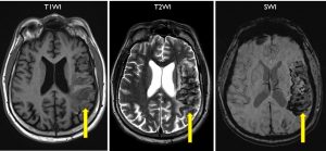
- Abnormal signal intensity seen in the left temporoparietal lobe sparing the parasagittal region (yellow arrows)
- It is heterogenously hypointense on T1/T2/FLAIR with gyriform hyperintense region seen
- Blooming artifact seen on SWI sequence
- Associated volume loss is seen
- These changes correspond to subacute MCA territorial infarction with hemorrhagic transformation
- Another abnormality seen as hyperintensity inferior to left lentiform nucleus to the left cerebral peduncle (red arrows) corresponding to changes along the corticospinal tract.
Radiological diagnosis: Wallerian degeneration due to massive infarction.
Discussion:
- Wallerian degeneration is disruption of the myelin and axons along the entire length of the nerve below the site of the lesion.
- It may result following neuronal loss due to cerebral infarction, trauma, necrosis, focal demyelination or hemorrhage.
- In post infarction, WD appears in the chronic phase (>30 days)
- CT is less sensitive than MRI the evaluation of this changes
- It is seen as contiguous tract of gliosis from a region of cortical or subcortical neuronal injury towards deep cerebral structures along the expected topographical course of the involved white matter tract
- The tract descend through subcortical white matter and form the anterior two-thirds of the posterior limb of the internal capsule. Then it pass through the ventral midbrain and continue through the pons. In the medulla oblongata corticospinal fibres collect into a discrete bundle forming the pyramid.
- The pyramid is a discrete triangular column on the ventral medulla oblongata next to the midline. This is why the corticospinal tract is also called the pyramidal tract.
