
Clinical:
- A 34 years old man
- Left eye blindness x 1 year
- Recent left eye proptosis
- No headache, seizure or limb weakness
- CT brain done showed left nasal mass (in other hospital)
- MRI done for further assessment of lesion
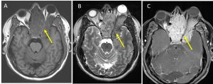
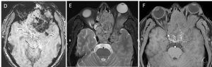
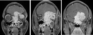
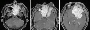
MRI findings:
- Large heterogenous enhancing mass (yellow arrows) within left sinonasal region measuring 7.5 (CC) x 6.8 (width) x 8 (AP) cm
- It extends into left anterior cranial fossa and into the extraconal space of the posterolateral aspect of the left orbit.
- Areas which exhibit blooming artifacts on SWI indicate persence of calcification within.
- Mass obliterates the left superior turbinate within nasal cavity and compresses the middle turbinate. The left osteomeatal complex is obliterated with mucosal thickening and secretion retention in left maxillary sinus. Mild extension through the meatal complex into the left maxillary sinus noted.
- Intracranial portion of left optic nerve compressed by the mass as well as in the orbital apex. Intraorbital left optic nerve displaced inferiorly. Left globe is proptosed.
- Not much inflammatory changes/oedema in the left orbit.
HPE findings:
- Macroscopy: specimen labelled as left nasal cavity mass
- Microscopy: section shows fragments of tissue infiltrated by sheets of tumor cells displaying oval to spindle-shaped, hyperchromatic and moderately pleomorphic nuclei with inconspicuous nucleoli and indistinct cytoplasmic borders. Multiple elongated and dilated vascular channels lined by single layer of endothelium noted. Foci of bone fragments and calcification noted. No necrosis present. Mitotic count is 3/10hpf.
- Immunohistochemistry shows tumor cells are:
- Positive for: Vimentin, Ki-67 (10%)
- Negative for: LCA, CK, CD99, S100, Synaptophysin, Chromogranin, EMA, HMB45, CD34
Diagnosis: Sinonasal haemangiopericytoma with atypical features.
Discussion (Sinonasal haemangiopericytoma):
- Hemangiopericytoma is an uncommon mesenchymal tumor, accounting for 1% of all blood vessel-related neoplasms and approximately 3% of all soft tissue sarcomas.
- It has a predilection for the long bones, pelvis and scapula; but, 15% occur in head and neck region, incidence of sinonasal HPC is less than 1%.
- It can occur in any age group and no sex predilection
- Radiographic features vary drastically and may resemble a malignant bone lesion. Radiographically, the lesion may be well circumscribed and sometimes appears to be radiopaque with displacement of surrounding structures.
Acknowldegement:
- Prof. Ahmad Sobri Muda
- Assoc. Prof. Dr Shahizon Azura Mohamed Mukari
Recent Comments