Case contribution: Dr Radhiana Hassan
Clinical:
- A 70 years old man
- On treatment for high uric acid for more than 20 years
- Complaint of painful swelling of left foot
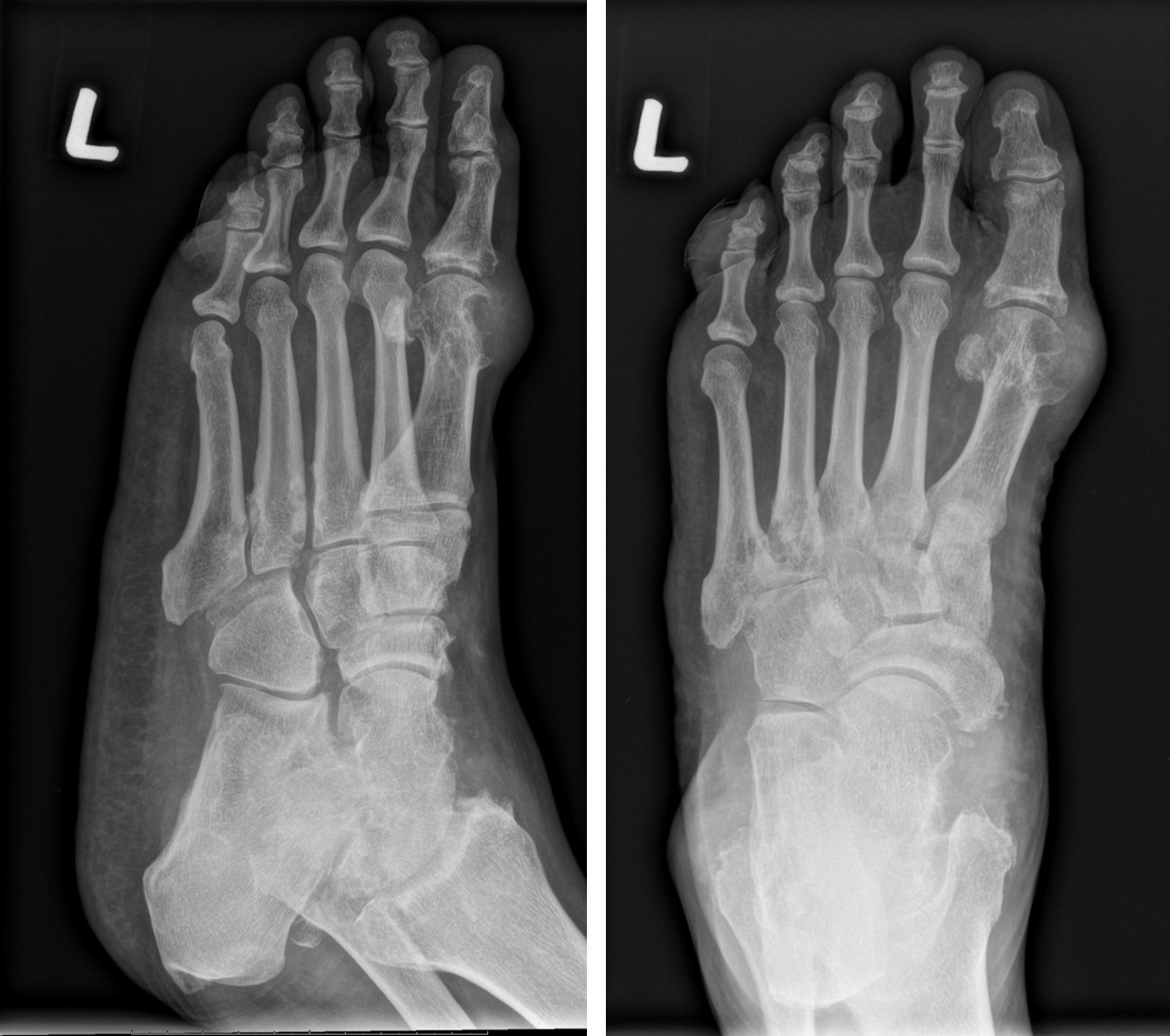
Radiographic findings:
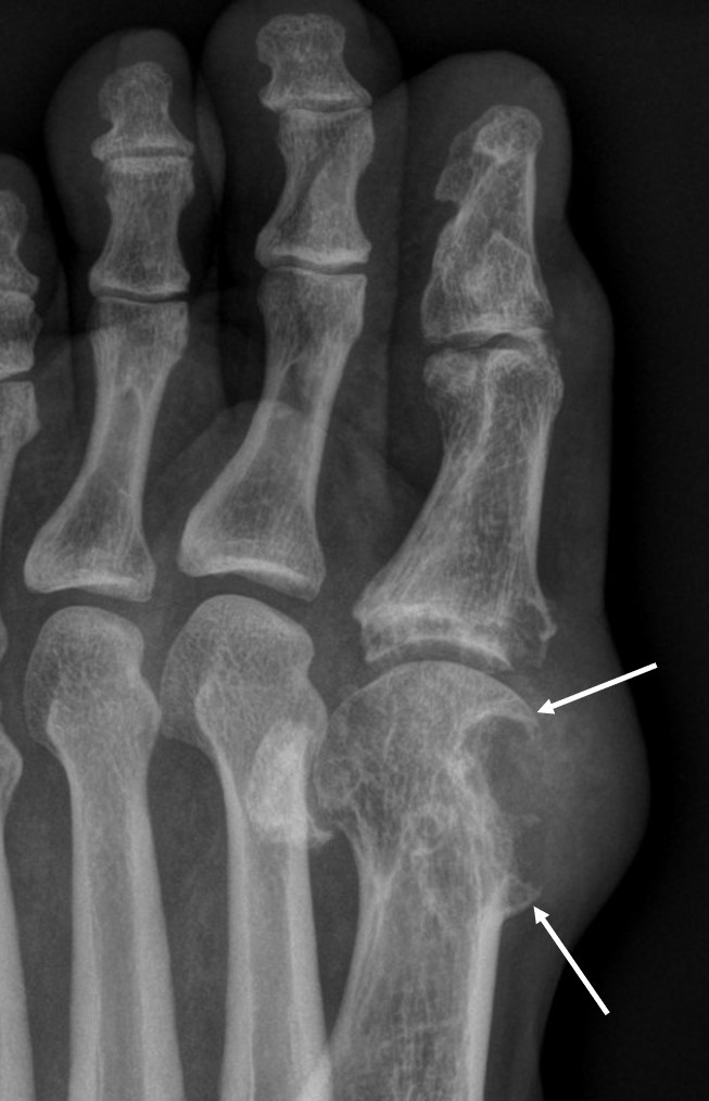
- Lytic lesions with well-demarcated margin at juxta articular region of first metatarsal and proximal phalanx (punch-out lesions)
- The bone lesions show sclerotic margin and overhanging edges (white arrows)
- There is associated oft tissue swelling with heterogenous opacity within in keeping with tophi
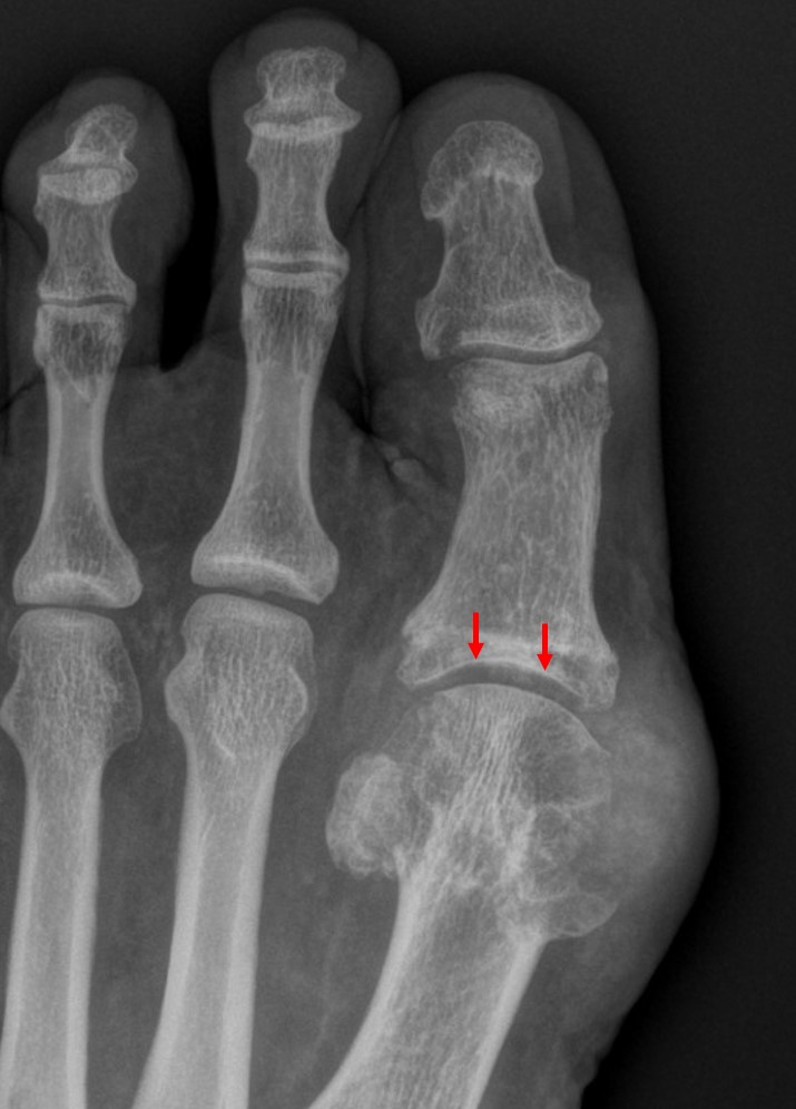
- The first metatarsophalangeal joint space (red arrows) is preserved
Diagnosis: Gouty arthritis
Discussion:
- Gouty arthritis is caused by deposition of sodium urate monohydrate crystals in synovial membrane, articular cartilage, ligaments and bursae leading to destruction of cartilage
- Age of onset usually more than 40 years
- Males much more often than females
- Stages:
- asymptomatic hyperuricemia
- Acute monoartiular gout
- Polyarticular gout
- Chronic multiple tophaceous gout
- Location: Joints: hands + feet (1st MTP most commonly affected), elbow, wrist, carpometacarpal compartment, knee, shoulder, hip, sacro-iliac joint (15%, unilateral), ear pinna > bones, tendon, bursa
- The early radiological signs of gout are joint effusion and periarticular edema.
- Radiographic examination eventually reveals a classic ‘punched-out’ lytic lesion with an associated overhanging edge at the distal metatarsals. Multiple marginal erosions and decreased joint space are seen at several metacarpal-phalangeal joints. These erosions contain sclerotic borders.
- The tophus, the hallmark of chronic gout, is a soft tissue nodule representing the body’s granulomatous immune reaction
- Osteopenia and the loss of joint space are usually not seen until advanced disease stages.
- The advanced stage is also characterized by joint destruction and severe deformities. Proliferative osseous change, intraosseous cysts, chondrocalcinosis (menisci, articular cartilate of knee), and olecranon bursitis can occasionally be seen in the patients with gout.
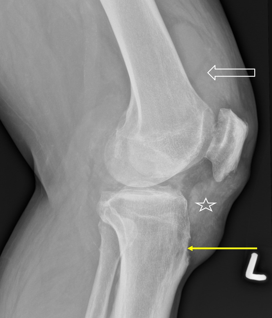
Progress of patient:
- Patient continues medical treatment
- Also had gouty arthritic changes involving the knee joint
- joint effusion (block arrow), tophi (star) and punch out lesion (yellow arrow)
Recent Comments