Case contribution: Dr Radhiana Hassan
Clinical:
- A 67 years old man with sudden onset of altered sensorium
- History of reduced oral intake and shortness of breath few days before
- Patient had underlying lung carcinoma, opted for conservative treatment
- History of left eye MALT lymphoma, completed radiotherapy
- GCS on arrival at ED=E4V1M5
CT scan findings:
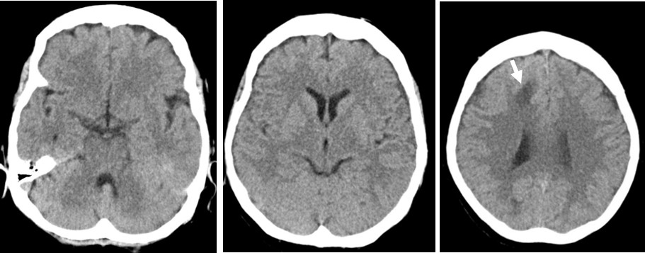
- CT scan show ill-defined hypodensity at right frontal lobe (white arrow) which show no enhancement on post contrast image
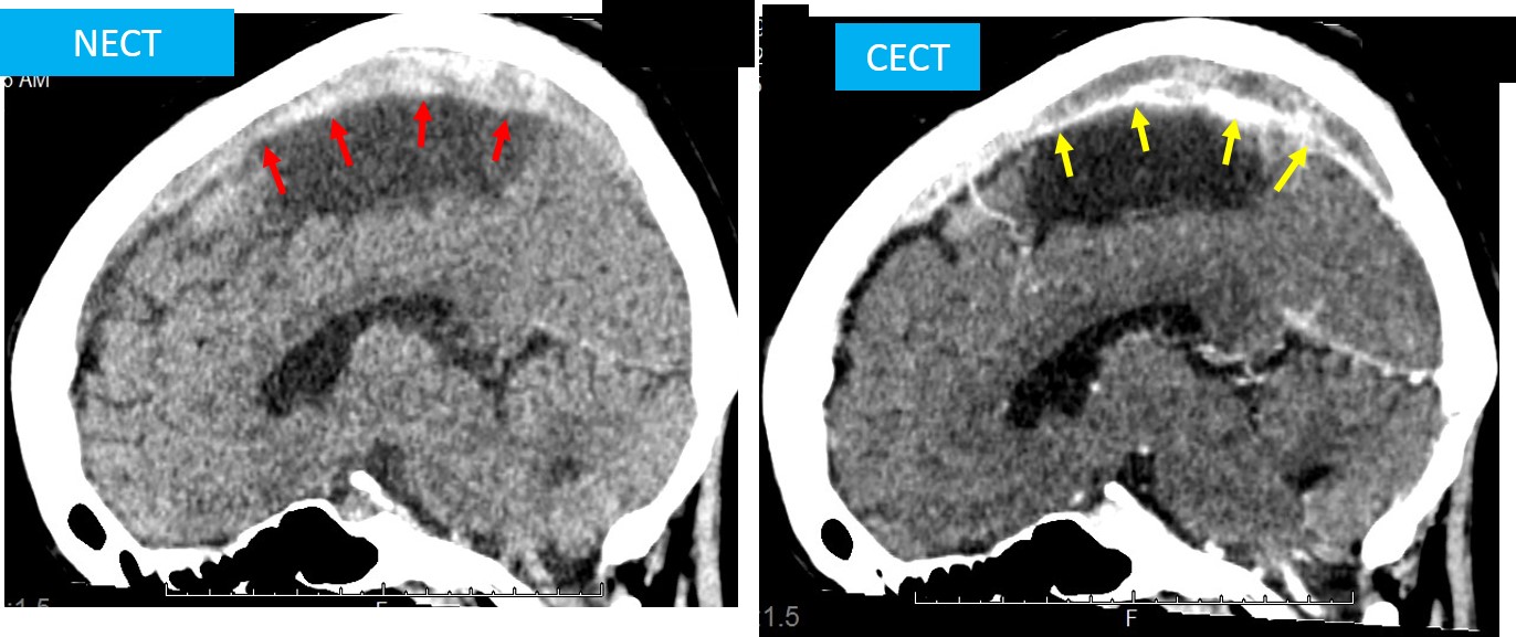
- Non-contrast reformatted sagittal image showed hyperdensity conforming to superior sagittal sinues (red arrows) seen. On post contrast images, filling defect (yellow arrows) are seen at the same region.
Diagnosis: Cerebral infarction caused by superior sagittal sinus thrombosis
Discussion (dural venous sinus thrombosis):
- Location: superior sagittal>transverse >sigmoid>straight sinus
- NCCT usually subtle findings include hyperdense clotted blood in the sinus, compression of lateral ventricles, infarction or edema, parenchymal hemorrhage and subdural collection
- CECT: filling defect in the sinus, enlargement of thrombosed veins near obstruction, shaggy irregular contour of the veins, gyral enhancement in periphery of infarction in 30-40% of cases, intense tentorial enhancement due to collaterals and dense transcortical medullary veins
- MRI can look for thrombus which show different signal intensity based on duration of thrombus, subcortical haemorrhagic infarctions, wall enhancement of thrombosed sinus, MRV show absence of flow
- Angiogram: non-filling thrombosed sinus, filling of cortical veins, deep venous system and cavernous sinus, parasagittal hemorrhages.
- Prognosis: high mortality
- Treatment: heparin, full recovery in 70% of cases
Progress of patient:
- Patient admitted and treated for hypovolaemia and hyponatraemia
- Had one episode of acute coronary symptoms while warded
- Died 5 days after CT scan
Recent Comments