Clinical:
- A 27 years old lady
- Presented with abdominal distension for few months
- No change in bowel habit
- Menstrual cycle normal
- Per abdomen showed a central mass
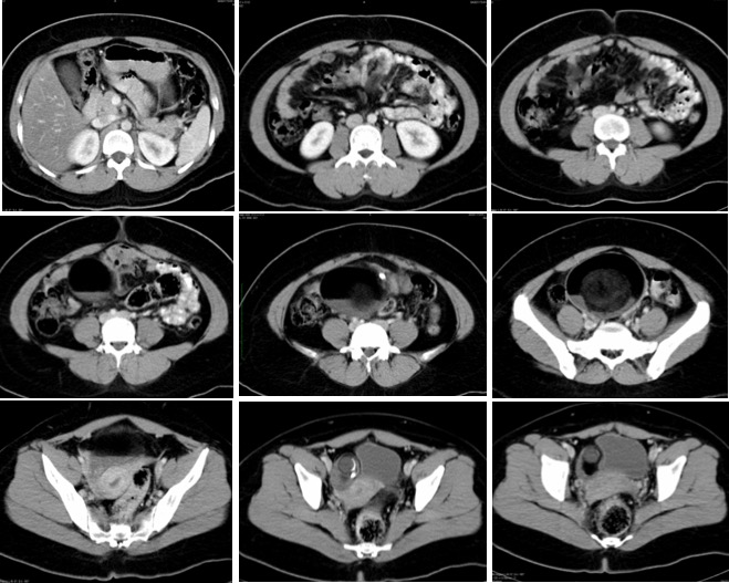
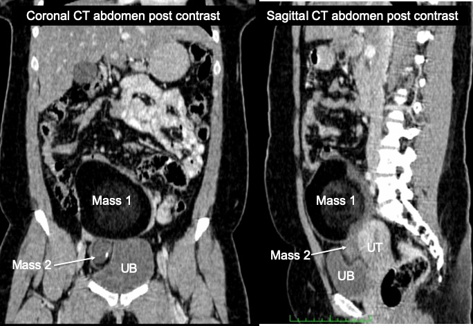
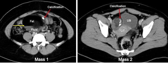
CT findings:
- Two pelvic lesions are seen (Mass 1 and Mass 2)
- The lesions have mixed density with wall calcification (red arrows)
- Fat fluid levels within the larger mass (yellow arrow)
- Presence of solid component which do not enhance after contrast
- Compression effect to the uterus and urinary bladder with clear plane of demarcation
- Displacement of bowel loops to the periphery
- No focal lesion in the liver, spleen, pancreas
- No enlargement of abdominal or pelvic nodes
- No free fluid
- UT= uterus, UB=urinary bladder
Intraoperative findings:
- Left ovarian mass measuring 18×12 cm in size, cystectomy done
- Right ovarian cyst measuring about 4×3 cm, ruptured during manipulation contains sebum and hair, cystectomy done
- Both fallopian tubes are normal
- Uterus normal
- No peritoneal seedling
Histopathological result:
- Macroscopy: specimen labeled as right ovarian cyst measuring 55x25x20mm. Cut section show a multiloculated cyst measuring 5-35 mm in diameter filled with yellowish fluid admixed with hair. The solid area measuring 10x5x5 mm. The cyst wall is 1-3mm in thickness. Another specimen labelled as left ovarian cyst consist of a lobulated mass measuring 120x90x40 mm. Cut section show uniloculated cyst filled with cheesy material admixed with hair. The cyst wall measures about 1-2 mm in thickness. No solid area.
- Microscopy: Right ovarian tumour consist of ectodermal and mesodermal tissue. The ectoderm is composed of stratified squamous epithelium with underlying sebaceous glands, hair, follicles and hair shaft. The mesoderm composed of glial tissue, chondroid tissue and bone. No immature element or malignancy seen. Section of left ovarian tumous show ovarian cyst wall lined by skin with underlying skin appendages. In areas the epithelium is replaced. No immature element or malignancy seen
- Diagnosis: Bilateral mature cystic ovarian teratoma
Final diagnosis: Bilateral mature cystic ovarian teratoma
Discussion:
- Ovarian mature cystic teratoma also known as dermoid cyst
- It is the most common ovarian neoplasm account for about 10-20% of all ovarian neoplasms.
- commonly seen in young women, typically around the age of 30 years
- They are bilateral in 10-15% of cases
- Ultrasound is the preferred imaging modality, typically seen as a cystic adnexal mass with some mural components. Most lesions are unilocular.
- Ultrasound is the preferred imaging modality. Typically an ovarian dermoid is seen as a cystic adnexal mass with some mural components. Most lesions are unilocular.
- As seen in this case, CT images typicaly demonstrate fat (areas with very low Hounsfield values),fat-fluid level and calcification.
- Rokitansky protuberance and tufts of hair can also be seen.
- The presence of most of the above tissues is diagnostic of ovarian cystic teratomas in 98% of cases
Recent Comments