Case contribution: Dr Radhiana Hassan
Clinical:
- A 40 years old lady
- Para 4
- Presented with back pain for one month
- Clinical examination is unremarkable
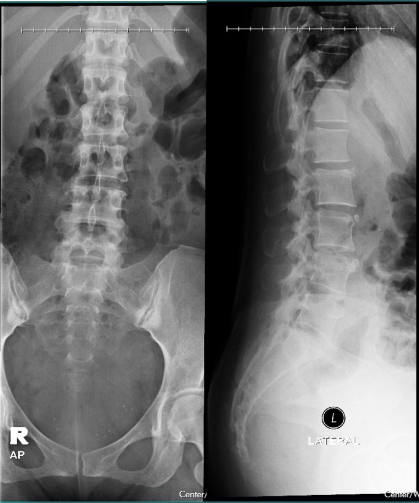
Radiographic findings:
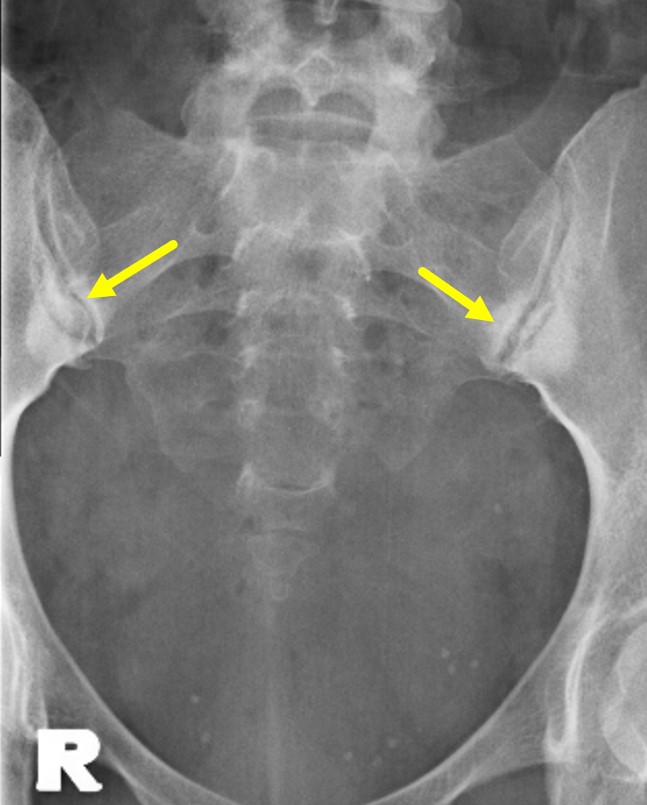
- Bilateral almost symmetrical sclerotic changes at both sacro-iliac joints (yellow arrows)
- Triangular in shape and more towards iliac side
- No narrowing of the SI joint spaces
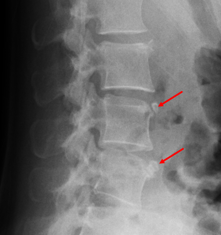
- Well-corticated opacities at anterosuperior corner of L3 and L4 vertebrae (red arrows)
- No reduction in vertebral body heights and intervertebral disc spaces
- No soft tissue mass
Diagnosis: Osteitis condensans ilii and Limbus vertebrae
Discussion:
- Osteitis condensans ilii is characterised by benign sclerosis of the ilium adjacent to the sacroiliac joint, typically bilateral and triangular in shape.
- It is usually asymptomatic but may cause back pain in about 1-2.5%
- The underlying aetiology is believed to be mechanical stress across the sacroiliac joint. That it is most often seen in women who have given birth supports this hypothesis; however, men and nulliparous women can be affected
- Lack of sacral involvement or joint space narrowing is considered diagnostic and thus, may obviate the need for further imaging.
- Unilateral disease has been reported.
- It carries a benign prognosis and may even resolve spontaneously.
- The main differential diagnosis is a sacroiliatis but as demonstrated in this case, the SI joint itself is normal, with no irregularity, erosions, or loss of joint space which are expected in sacroiliatis.
- Limbus vertebra is a well-corticated unfused ossification centre
- Anterior limbus vertebrae are generally asymptomatic and are detected incidentally.
- Posterior limbus vertebrae are far less common but have been reported to cause nerve compression.
Recent Comments