
Clinical:
- A 20 years old lady
- Diagnosed multiple sclerosis 6 years ago
- Initial presentation: bilateral spastic paraparesis
- Also had bilateral cerebellar sign and unsteady gait.
MRI findings:
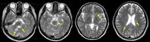
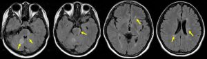
- There are multiple T2-hyperintense lesions in the cerebellum, midbrain, and periventricular region (yellow arrows): dissemination in space
- The lesions at periventricular region are perpendicularly orientated to the ventricle
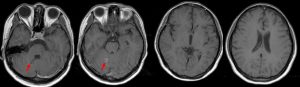
- Some of the lesions show enhancement post contrast (red arrows) while others show no enhancement: dissemination in time
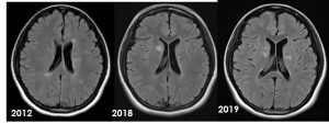
- new lesions are seen during subsequent imaging
Diagnosis: Multiple sclerosis
Discussion:
- Multiple sclerosis is an acquired chronic relapsing demyelinating disease involving the central nervous system
- The presentation is usually between adolescence and the sixth decade, with a peak at approximately 35 years of age
- Revision of McDonald diagnostic criteria as follows:
| 2005 | 2010 | 2017 |
| Gado-enhancing lesion or 9 T2-lesions | Periventricular | Periventricular |
| At least 3 periventricular T2-lesions | Juxtacortical | Juxtacortical/Cortical |
| At least 1 infratentorial or spinal cord T2-lesion | Infratentorial | Infratentorial |
| At least 1 juxtacortical T2-lesion | Spinal cord (excluded if symptomatic lesions) | Spinal cord |
| (at least 3 of above required) | 1≥ T2 lesion at least 2 of 4 areas | 1≥ T2 lesion at least 2 of 4 areas
No distinction of symptomatic or non-symptomatic lesion |
Recent Comments