
Clinical:
- A 41 years old man
- Presented with left pulsatile tinnitus for 2 years
- Associated with left ear blockage and reduced hearing
- No history of ear discharge, no vertigo
- Clinical examination showed reddish mass at left ear, pulsatile, medial to TM,
- TM is intact. Right ear is normal.
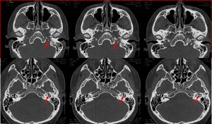
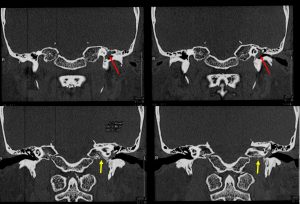
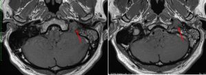
HRCT temporal bone and MRI findings:
- soft tissue mass in the left jugular foramen with mild enhancement post contrast (red arrows)
- erosion of left jugular foramen also seen (yellow arrows)
- mass with similar signal intensity seen in the left middle ear, abutting the tympanic membrane
- these two masses seems to be in continuity or represent same mass with extension to the region
- left ossicles are normal
- left mastoiditis seen
- no intracranial extension, right ear is normal
Diagnosis: left glomus jugulotympanicum tumour
Discussion:
- glomus jugulotympanicum is a glomus jugulare paraganglioma that has spread superiorly to involve the middle ear cavity
- seen as mass in jugular foramen with permeative destructive changes along the superolateral margin of jugular foramen
- CT is useful to assess bony margins of tumour which are typically showed erosion or moth-eaten appearance
- CT also good to assess integrity of ossicles and bony labyrinth
- MRI: Hypo hyper T1/T2 and intense enhancement. Salt pepper appearance from blood products
Recent Comments