Clinical:
- A 53 years old lady
- Presented with abdominal distension for few months
- No constitutional symptoms
CT scan findings:
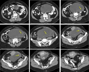
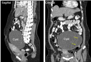
- A cystic mass measuring 17x15x9 cm
- It has thin wall with thin septae seen (yellow arrows)
- No enhancement, no calcification and no fat component
- No ascites, no surrounding fat streakiness.
- No local infiltration, no distant metastasis
HPE findings:
- Macroscopy: specimen labelled as ovarian cyst, uterus, cervix and fallopian tubes. The ovarian cyst measures 185x170x110 mm. The whole specimen weighs 1750 gms. The ovarian cyst has smooth and intact surface. Cut section shows a multiloculated cyst filled with serous fluid. The cyst wall measures from 1mm to 4 mm thick. The cysts ranges from 15-110 mm. No solid area seen.
- Microscopy: sections show a benign cystic tumour and the cyst wall lined by a single layer of mucinous epithelium. The cells are basally located and benign looking nuclei with abundant mucin containing cytoplasm. No borderline feature or evidence of malignancy noted.
Diagnosis: Left ovarian mucinous cystadenoma
Discussion (Ovarian mucinous cystadenoma):
- Mucinous cystadenoma is a benign epithelial tumour with peak incidence around 30-50 years of age.
- Mucinous cystadenoma typically presents as a unilateral, multilocular, voluminous, cystic, ovarian lesion. Macroscopically, the content is mucoid. A benign mucinous cystadenoma has no solid portion. Its wall is smooth but may sometimes have a few papillary projections.
- Ultrasound shows typically large cystic adnexal mass, multilocular with numerous thin septations. The loculations may contain low-level internal echogenicity due to increased mucin content (different locules may contain various degrees of echogenicity)
- On MRI benign mucinous cystadenoma typically presents as a large, multilocular, cystic, ovarian lesion. Some loculi are spontaneously hyperintense on T1-weighted MR images (related to the variable concentration of mucin in the loculi). These loculi are more or less grouped together sometimes giving a “honeycomb” or “stained glass” pattern.
- The criterion indicating a benign lesion is the absence of areas of tissue (the absence of irregular septa or solid portions).
Recent Comments