
Clinical:
- A 23 years old lady
- Presented with abdominal distension
- Gradually increase in size
- No constitutional symptoms
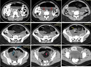
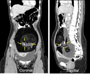
CT scan findings:
- A large lesion measuring 16x18x10 cm, mostly probably arising from the left ovary
- Heterogenous density, thin wall with no enhancement
- Presence of intralesional calcification (blue arrows)
- Presence of fat components (star)
- Presence of fat-fluid levels (red arrows)
- A soft tissue density projecting into the cyst is seen (yellow arrows) in keeping with Rokitansky nodule
- Floating ball sign (white arrows)
- No ascites, no local infiltration, no distant metastasis
Intra-operative findings:
- Left ovarian tumour size 15x10x10 cm
- Cystic, freely mobile, tumour exteriorised, closer inspection revealed hair strands within the tumour
- Right ovary and both fallopian tubes are normal and freely mobile
- Mild adhension between the descending colon and left pelvic side wall
- Omentum, bowel, liver and under surface of diaphragm is normal.
- Right cystectomy and adhesiolysis done
HPE findings:
- Macroscopy: specimen labelled as ovarian tumour consists of an ovarian tumour measuring 180x140x80 mm. The whole specimen weighs 700 gms. The external surface is smooth and is partially ruptured. Cut section shows a unilocular cyst containing cheesy material admixed with hair. The cyst wall measures 1 to 3 mm thick. There are 2 solid area seen. The larger one is 50x40x10 mm and the smaller one measures 40x38x18 mm. There are two teeth on the smaller solid area. The largest solid area measures 55x45x20 mm. Cut section shows yellowish cut surface.
- Microscopy: section shows an ovarian cyst wall lined by flattened, cuboidal epithelium in the thinner areas. The solid areas are covered by stratifying squamous epithelium with underlying sebaceous glands, hair follicles, blood vessels, foci of lymphoid tissue and smooth muscle bundles. There is no immature or malignant tissue seen.
Diagnosis: Mature cystic teratoma of left ovary
Discussion (Rokitansky nodule):
- A Rokitansky nodule is also known as dermoid plug.
- It refers to a solid protuberance projecting from an ovarian cyst. It often contains calcific, dental, adipose, hair, and/or sebaceous components.
- It is quite common in mature cystic teratoma, in some series reported in 98% of cases.
- It might be seen as a focal mural thickening, round structure protruding into the cystic lumen, a bridging tissue across the cyst, a cystic structure or sometimes only tooth.
- It ranges from 10 to 45 mm.
- This nodule has the highest propensity to undergo malignant transformation.
- Features suspicious of malignant transformation include contrast enhancement, an obtuse angle between the soft tissue and the wall of the cyst and extracapsular tumour growth with extension into the adjacent structures.
Recent Comments