
Clinical:
- A 10 years old girl
- Presented with pain over right leg
- Aggravated by prolong walking and standing
- No history of trauma or fever
- Partially relieves with analgesic
- No constitutional symptoms
- Clinically a swelling felt at proximal tibia, 2×2 cm, bony hard, non-tender, no overlying skin changes. Range of motion right knee is normal. No neurological deficit.
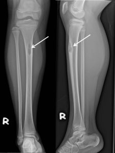
Radiographic findings:
- A lytic lesion at anterior cortex of proximal right tibia
- It measures about 3.1 x 0.8 cm.
- Narrow zone of transition
- Presence of endosteal scalloping
- No matrix calcification
- No cortical breach , periosteal reaction or associated soft tissue mass is seen.
- Knee and ankle joints are intact.
Diagnosis: Non ossifying fibroma of right tibia
Discussion:
- Non ossifying fibroma is a common focal lesion in the bones.
- It is estimated to be present in up to 30% of the asymptomatic population in the first and second decade of life
- Most common between 8-20 years of age
- They are not neoplasms and, according to WHO, belong to the group of developmental abnormalities
- On radiograph seen as lytic lesion, usually oval, surrounded with a thin sclerotic rim, with a long axis parallel to the axis of the bone
- The lesions are usually found in the metaphyseal areas, mainly in bones constituting the knee joint
- Geographical, sclerotic rim, endosteal scalloping
- Migrates toward centre of diaphysis and resolve with age
- May undergo pathologic fracture, no malignant transformation
Progress of patient:
- On conservative management with analgesic
- Repeat radiograph six months after initial presentation shows no interval change of bone lesion
Recent Comments