")
Clinical:
- A 22 years old lady
- Sudden onset of left sided weakness for 2 weeks
- Left sided loss of sensation since 13 days.
- Clinically had loss of left nasolabial fold
MRI findings:
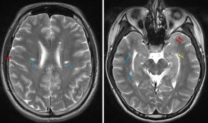
- There are multiple white matter lesions
- Juxtacortical lesions involving U-fibres (red arrows)
- Periventricular lesions (blue arrows)
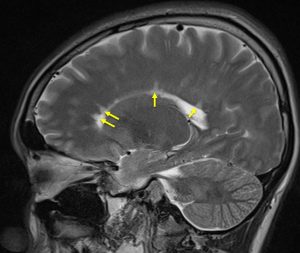
- Dawson fingers (yellow arrows)
- These lesions high signal intensity on T2 and FLAIR sequences, not restricted on DWI and not enhanced post contrast.
Radiological diagnosis: Consistent with multiple sclerosis
Discussion (Dawson fingers):
- Initially described by James Dawson on histopathological specimens in MS as “wedge-shaped areas with broad base to the ventricle, and extensions into adjoining tissue in the form of finger-like processes or ampullae, in each of which a central vessel could usually be found”.
- On MRI, Dawson’s fingers are described as elongated, flame-shaped, hyperintense lesions best seen on sagittal FLAIR images (as shown in this case). They are oriented along subependymal veins and thus are perpendicular to the walls of lateral ventricles.
- This finding is not found in MRI of Neuromyelitis Optica Spectrum Disorders (NMOSD) and can be a differentiating features from MS.
Recent Comments