
Clinical:
- A 57 years old lady
- Fall with outstretched hand
- Complains of painful swelling of the right wrist
- Clinically swollen and deformed distal right upper limb, at wrist region
- Shoulder and elbows are normal
Radiographic findings:
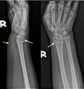
- A transverse fracture at distal radius (white arrows)
- No extension to involve the articular surface
- There is dorsal angulation of fracture fragment
- Distal radio-ulnar joint is normal
- No fracture of ulnar styloid or the carpal bones.
- There is associated soft tissue swelling anteriorly (yellow arrow)
Diagnosis: Colles fracture
Discussion:
- Colles fracture is an extra-articular fractures of the distal radius
- They consist of a fracture of the distal radial metaphyseal region with dorsal angulation and impaction, but without the involvement of the articular surface.
- This is the most common type of distal radius fracture.
- An associated ulnar styloid fracture is seen in about 50% of cases, which is not seen in this case.
- The majority are treated with closed reduction and immobilization.
- Complications include malunion causing dinner fork deformity, secondary osteoarthritis, median nerve palsy and post traumatic carpal tunnel syndrome
Recent Comments