
Clinical:
- A 23-year old lady
- No known medical problem
- Presented with chronic headache.
Imaging findings:
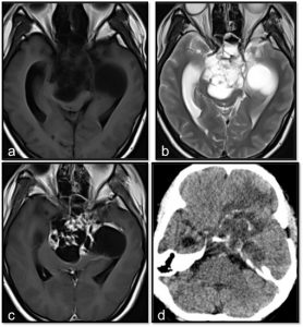
- Axial MRI images in (a) T1-weighted, (b) T2-weighted and (c) T1-post contrast
- show a lobulated multicystic lesion at sellar and suprasellar region with minimal solid component that show enhancement after contrast administration.
- Non-contrast axial CT scan (d) show presence of intralesional calcifications.
Diagnosis: Craniopharyngioma (adamantinomatous type-HPE proven)
Discussion:
- Craniopharyngiomas are benign neoplasms that typically arise in the sellar/suprasellar region.
- It account for 1-5% of primary brain tumours and can occur anywhere along the infundibulum to the pituitary gland.
- Craniopharyngioma are found in patients of all ages. Bimodal distribution seen with first peak at 10-14 years and second peak in adults aged over 50 years old.
- There are two pathological types; adamantinomatous and papillary type.
- Adamantinomatous type is more common (90% of cases) and seen in pediatric patients, whereas papillary type is less common (10% of cases) and occurs in adult patient.
- Imaging features depend on the type of tumour with a minority of cases having both adamantinomatous and papillary component and may show overlapping features.
- In majority of cases, the tumour have a large suprasellar component (95%), involving both supra and intrasellar (75%) and minority are purely suprasellar (20%).
- It may be associated with expansion of the pituitary fossa and larger tumours can extend in all directions, frequently distorting the optic chiasm or compressing the midbrain causing obstructive hydrocephalus.
- Adamantinomatous types shown in this case typically has a lobulated contour with multiple cystic lesions. Minimal solid components are present and this solid component enhance vividly both on CT and MRI.
- Overall, calcification is very common.
Recent Comments