
Clinical:
- A 50 years old lady
- No previous medical illness
- Presented with early satiety and progressive abdominal distension. No obstructive or constitutional symptoms.
CT scan findings:
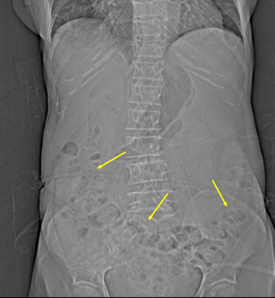
- scanogram shows displacement of bowel loops inferiorly (yellow arrows)
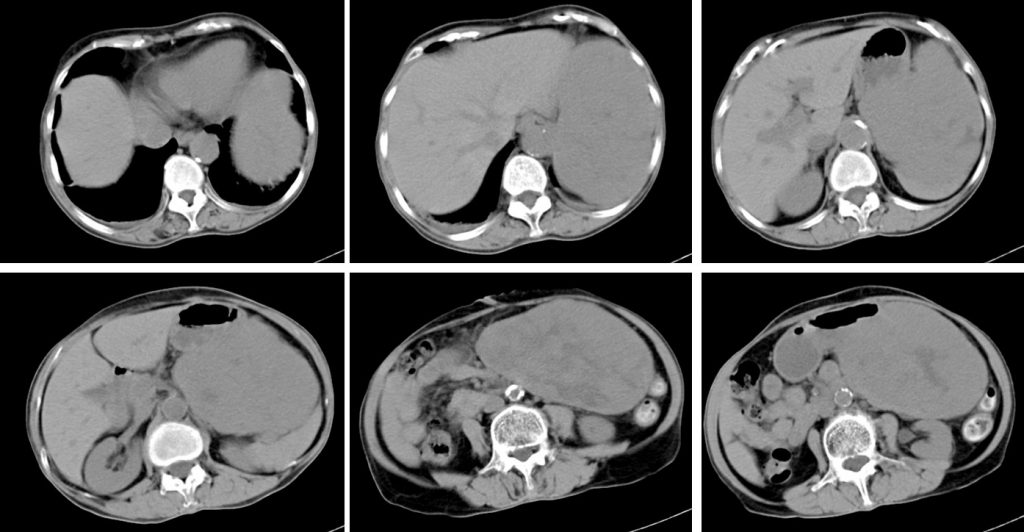
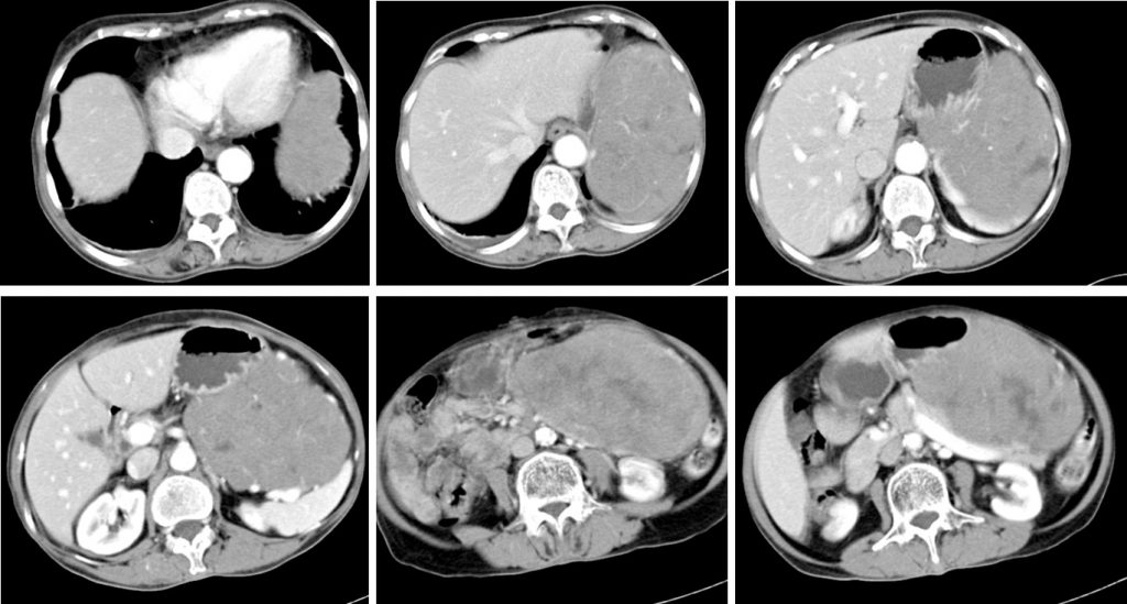
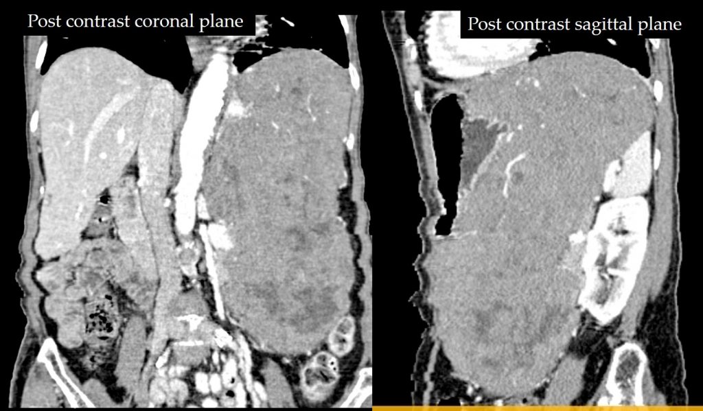
- CT scan shows a huge mass arising from posterior wall of stomach
- It shows heterogenous enhancement with areas of central necrosis
- No calcification is seen within the mass lesion.
- Compression and displacement of surrounding structures are seen, however clear fat plane is still preserved.
- No contrast extravasation to suggest active hemorrhage.
- No dilatation of bowel loops. No enlarged nodes
Diagnosis: Stomach GIST (HPE proven)
Discussion:
- Gastointestinal stromal tumours (GIST) are the most common mesenchymal tumors of the gastrointestinal tract.
- GIST accounts for about 5% of all sarcomas.
- Stomach is the commonest location for this tumour (about 70% of cases).
- Usually occur after 40 years of age, most seen in older patients
- In this case it bulges extramural not causing any bowel obstruction
- Imaging appearances vary with size and location.
- Typically the mass is of soft tissue density with central areas of necrosis.
- The tumors are often exophytic, Enhancement is typically peripheral (due to central necrosis)
- Calcification is uncommon (3%)
- Lymph node enlargement is not a feature
- Metastases (distant, peritoneal, omental) or direct invasion into adjacent organs may be seen in more aggressive lesions
Recent Comments