
Clinical:
- A 60 years old lady
- Post menopausal
- Presented with chronic constipation
- No other obstructive symptoms
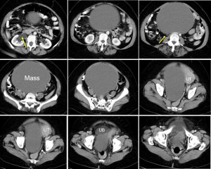
CT scan findings:
- There is a huge cystic mass measuring 14x18x19 cm
- It shows thin wall, no septation, no enhancement, no calcification and no fat component within the lesion
- Compression of right ureter causing right hydronephrosis (yellow arrows)
- Compression effect to the sigmoid colon (white arrows)
- No local infiltration to surrounding structures, no distant metastasis, no ascites
Intra-operative findings:
- Total abdominal hysterectomy, bilateral salpingo-oophorectomy, omentectomy and pelvic nodes dissection
- Right ovarian mass 28x20x15 cm, solid cystic lesion, adhered to right lateral pelvic wall and posterior abdominal wall, ruptured during dissection of tumour
- Left ovary adhered to the lateral wall of the uterus, uterus atrophic.
- Pelvic nodes not enlarged
HPE findings:
- Macroscopy: specimen consist of uterus, cervix, left fallopian tube, left ovary, right fallopian tube and right ovarian cyst. The right ovarian cyst measures 130x110x20 mm. The wall thickness is 1 to 6 mm. There are multiple fungating brownish brownish lesion seen at the inner surface of the ovarian cyst measuring 3 to 33 mm in diameter.
- Microscopy: Sections of the right ovarian cyst show it is lined by ciliated tubal-type epithelium. In areas there are papillae with complex branching without stromal invasion. No nuclear atypia, mitoses, haemorrhage or necrosis seen.
Diagnosis: Right ovarian cyst (borderline serous tumour)
Discussion:
- Ovarian tumors of borderline malignancy are a distinct histologic and clinical entity diagnosed in up to 15% of patients presenting with an ovarian neoplasm.
- They are common in younger women and often appear clinically to be benign.
- Compared with frankly malignant tumors, borderline tumors have a much better prognosis and, because they are noninvasive, may be treated less radically than invasive ovarian cancer.
- They are common in younger women and often appear clinically to be benign.
- On gross pathologic examination, they are unilocular or multilocular cystic tumors with or without epithelial proliferations on the outer tumor surface.
- About one third are bilateral.
Recent Comments