
Clinical:
- A 24 years old lady
- Progressive abdominal distension
- No obstructive symptom
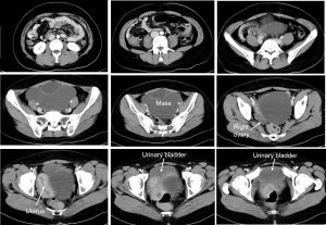
CT scan findings:
- A cystic mass in the left pelvic region measuring 15x12x13 cm. No solid component or papillary projection.
- It shows thin wall with multiple thin mildly enhancing septae
- There is no fat component or calcification within the mass lesion.
- The right ovary is normal. No ascites
- No local infiltration to surrounding structures
Intra-operative findings:
- Laparotomy and left cystectomy and release of large bowel adhesion
- Left ovarian cyst, 15×18 cm, multiloculated cystic, no solid component, posterior to uterus, no obvious surface deposits, mild adhesion between the descending colon and the ovarian cysts
- Both fallopian tubes are normal
HPE findings:
- Macroscopy: left ovarian cyst consist of a cystic mass measures 110x80x50 mm. Cut section shows multiloculated cyst filled with jelly-like material. The locules ranges from 5 mm to 50 mm in diameter. The wall thickness is about 1 mm. No solid area seen.
- Microscopy: sections show fibrocollagenous ovarian cyst wall and locules lined in areas by mucin-secreting, columnar epithelium. There is no tufting, multilayering, atypia or mitoses seen Foci of calcification are present in the wall.
- Diagnosis: left ovarian cyst: Mucinous cystadenoma
Diagnosis: Ovarian mucinous cystadenoma
Discussion:
- Mucinous cystadenomas of the ovary are known for their potential to grow to massive proportions.
- They are often incidentally diagnosed.
- They are typically benign tumours accounting for 15 percent of ovarian neoplasms and up to 80 percent of all mucinous tumors.
- Ovarian mucinous cystadenomas are characteristically unilateral, only 5 percent presenting bilaterally
- The peak incidence occurs among women who are between 30 and 50 years of age.
Recent Comments