Clinical:
- A 34 years old lady
- Background history of right congenital nystagmus with reduced vision and glaucoma of same eye.
- Presented with right temporal visual defect
- Normal intraoccular pressure diagnosed with VF test.
- MRI to rule out any space occupying lesion


MRI findings:
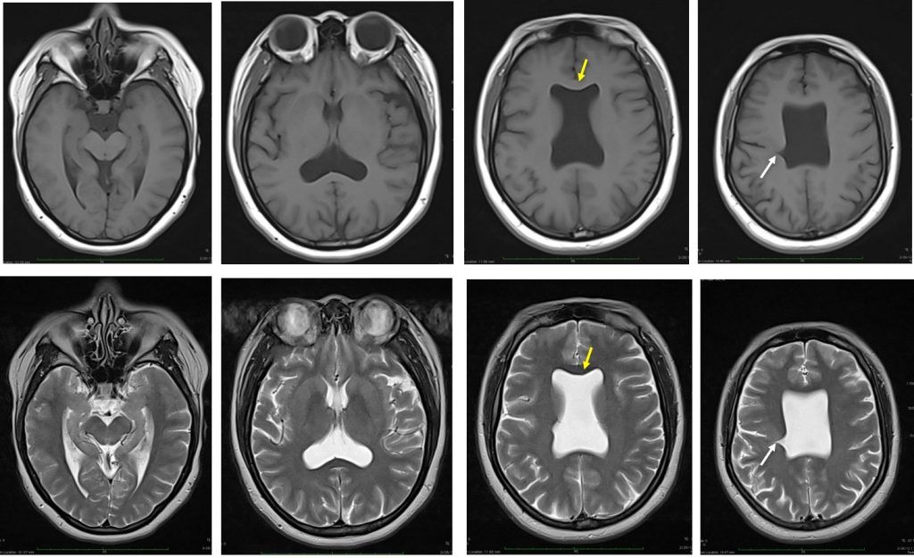
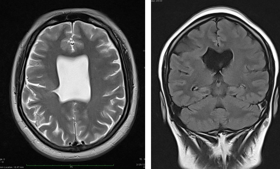
- Absent septum pellucidum giving rise to a box-like configuration of the frontal horns of the lateral ventricles (yellow arrows).
- There is a incomplete cleft from the lateral wall of the body of the right lateral ventricle which traverses across almost to join the sulci (white arrows). This incomplete cleft is lined by the grey matter.
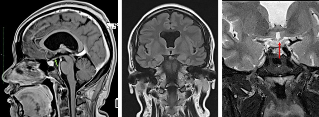
- The corpus callosum is intact.
- Both optic nerves and optic chiasm appear small (red arrow).
- The pituitary gland is of normal size but the pituitary stalk appears small (green arrow).
- Normal brain parenchyma signal intensity.
- Both orbits are grossly normal. The bilateral extraoccular muscles are normal. No intraorbital lesion.
Diagnosis: Septo-optic dysplasia with schizencephaly.
Discussion:
- Septo-optic dysplasia is a congenital disorder belonging to the mid-line brain malformations group.
- It can be highly variable resulting in a wide heterogeneity, however its classical triad includes the following:
- Optic nerve hypoplasia
- Agenesis of midline structures (namely septum pellucidum and corpus callosum)
- Hypoplasia of the hypothalamo-pituitary axis
- This disorder is associated with maternal diabetes, quinidine ingestion, antiseizure medication, drug abuse, cytomegalovirus and congenital brain malformations (Chiari II and aqueductal stenosis).
- Nearly 80% have pituitary dysfunction.
- Septo-optic dysplasia can also present with other specific brain abnormalities uch as schizencephaly as demonstrated in this case and called “SOD plus syndrome”.

Recent Comments