
Clinical:
- An 18 years old girl
- Underlying beta thalassemia major on frequent blood transfusion
- Presented with headache, fever and meningism.
- CT brain shows ventriculitis
- EVD inserted-pus drained.
MRI findings:
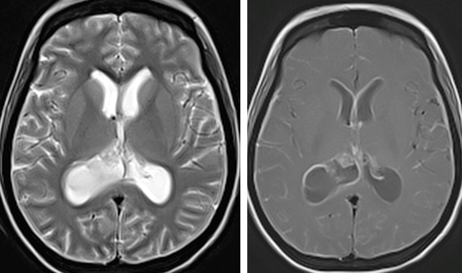
- There is a cystic lesion seen within the atrium of the left lateral ventricle with barely perceptible thin enhancing walls. It measures 2.9 x 2.8 x 2.9cm (APxWxCC).
- Layering signal intensity seen within suggestive of sediments. Sediments are also seen within the right lateral ventricle.
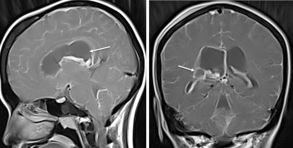
- The ventricles are dilated. The ependymal linings show marked enhancement post contrast.
- Generalised leptomeningeal enhancement noted in both cerebral sulci extending into the visualized upper cervical region.
- No abnormal signal intensity seen within the brain parenchyma.
Progress of patients:
- Patient was treated as intraventricular abscess.
- Condition worsened after one month of treatment
- EVD inserted twice, hydrocephalus increased in severity.
- First and second CSF sample shows no significant finding.
- Third CSF sample positive for TB
- Patient shows good response after antiTB treatment.
Repeat MRI prior to anti-TB treatment:
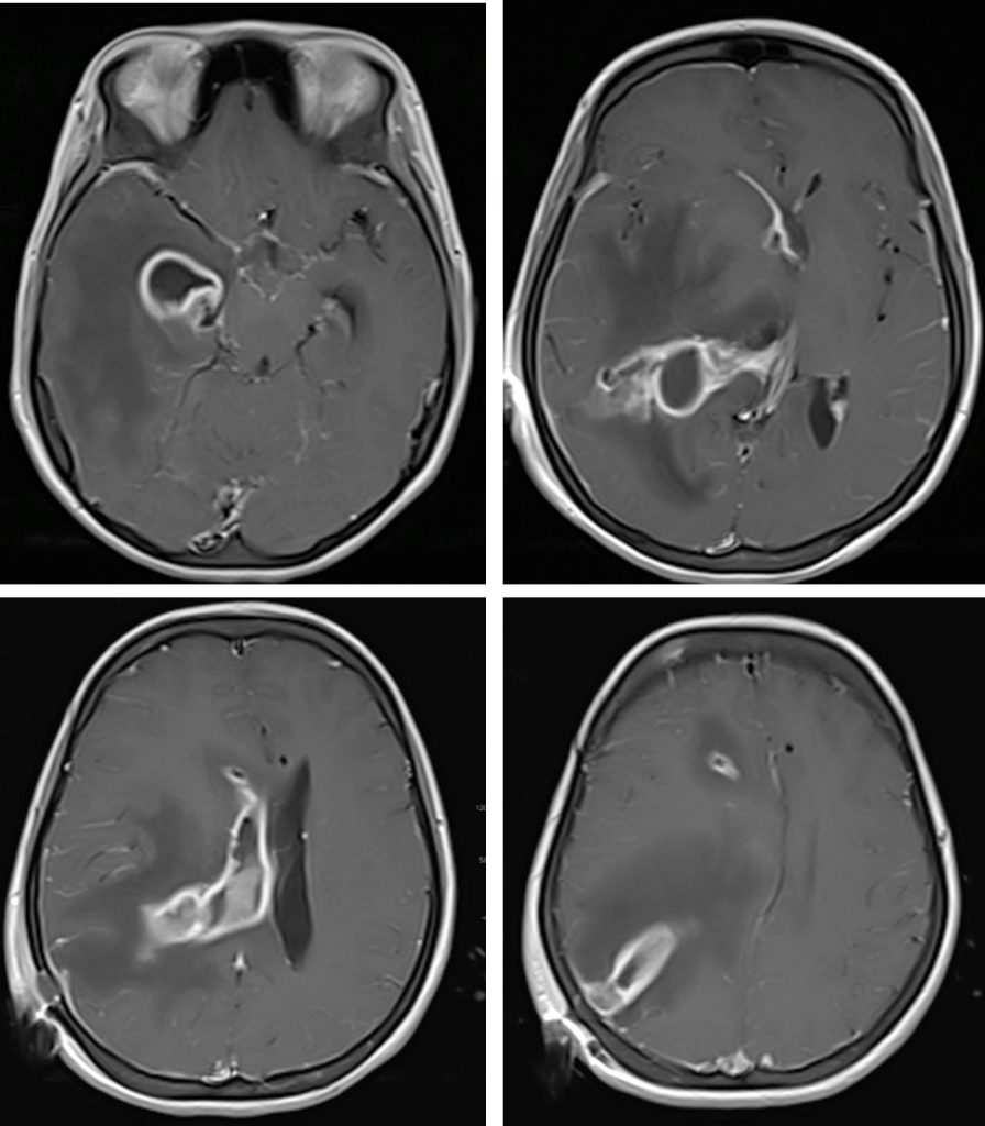
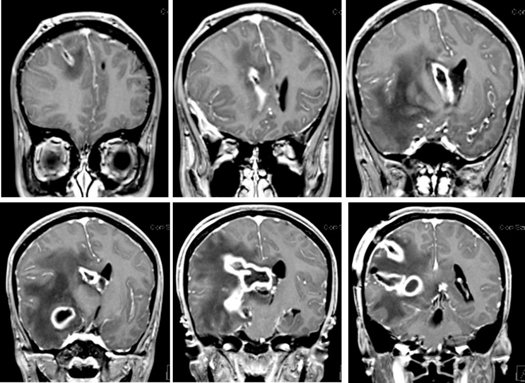
- Previously seen multiseptated collections involving entire right lateral ventricle are more enhancing with thickened wall.
- Presence of debries within the right temporal horn, pocket of air seen within as noted previously.
- The perilesional oedema more conspicious in this study compared to previous images.
- Enhancement along the EVD are more prominent .
- The leptomeningeal enhancement is more pronounced in this study.
Discussion:
- Ventriculitis refers to inflammation of the ependymal lining of the cerebral ventricles.
- Many causes can lead to ventriculitis, such as meningitis, cerebral abscess, trauma, post
brain procedure and secondary to brain neoplasms. - Tuberculous ventriculitis is a rare complication of the central nervous system tuberculosis.
- The imaging findings are abnormal meningeal enhancement predominant
in the basal cisterns, hydrocephalus, and enhancement of the ependymal
surface (granulomatous ependymitis) with intraventricular pus or debris. - Parenchymal abnormalities may be seen including masses of granulomatous
tissue (low signal on T2WI), granulomatous abscesses (hypo- or iso or
central hyperintensity with a hypointense rim on T2WI). - Vasculitis and ischaemic events can also occur.
Recent Comments