Clinical:
- A 53 years old man
- Underlying hypertension
- Presented with chronic cough and worsening shortness of breath
- Associated with constitutional symptoms.

Radiographic findings:
- There is total opacification of the left hemithorax causing non-visualization of left cardiac border and left hemidiaphragm. No air bronchogram sign within.
- There is displacement of trachea (yellow arrow) and mediastinum to the right side.
- There is occlusion of left main bronchus (white arrow).
- The aerated right lung is clear. No nodule or consolidation seen.
- No blunting of right costophrenic angle.
- No obvious bone destruction seen.
Radiological diagnosis: Opaque left hemithorax with mediastinal shift
Discussion:
- Whitening out of half of the lung field on a chest X-ray is known as opacification of a hemithorax, and its presence usually indicates a significant disease in a patient.
- Opaque hemithorax with mediastinal shift away from the opacified side can occur when there is increase of volume of affected hemithorax.
- The differential diagnosis include massive pleural effusion, diaphragmatic hernia, lung mass and diaphragmatic rupture.
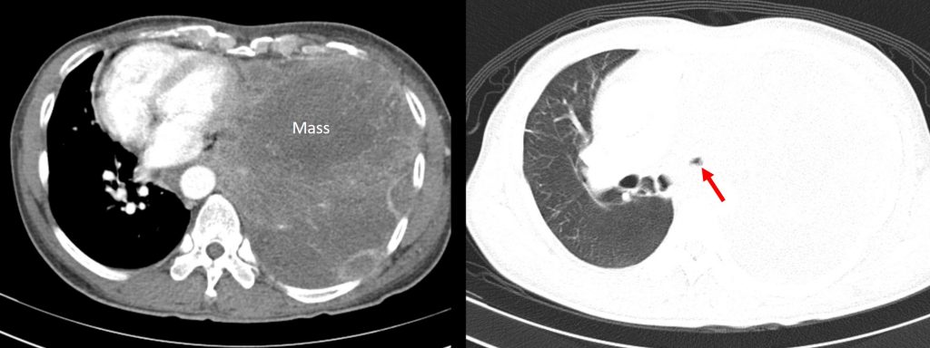
Progress of patient:
- CT scan performed showing huge mass in the left lung total obstruction of left main bronchus causing total collapsed of left lung.
- Biopsy was performed.
HPE findings:
- Macroscopy: specimen labelled as lung consists of multiple strips of brownish tissue measuring 2 to 25 mm in length.
- Microscopy: sections show multiple strips of lung tissue, fibrocollagenous tissue, skeletal muscle and a small bit of skin. The lung tissue and fibrocollagenous tissues are infiltrated by malignant tumour that is arranged as cord and clusters within fibrotic to desmoplastic stroma
- Immunohistochemical stains: CK7, CK20, TTF-1,p40 are negative. CD55 is positive. Chromogranin, synaptophysin negatives
Interpretation: lung biopsy; poorly differentiated carcinoma.
Recent Comments