
Clinical:
- A 45 years old male
- Presented with right upper limb weakness and headache.
- CT brain showed left fronto-parietal lesion with midline shift.
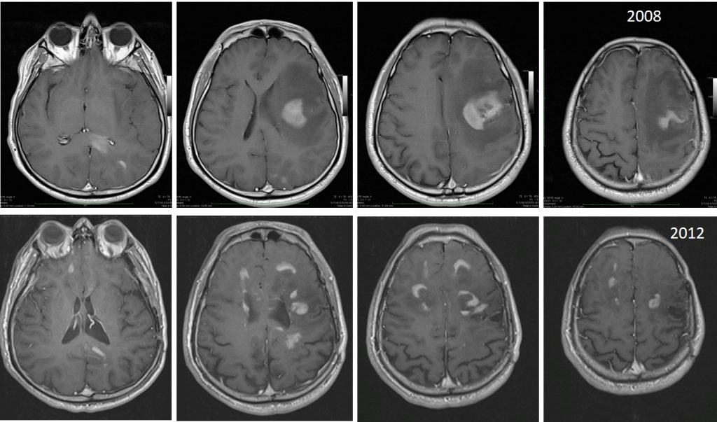
MRI findings:
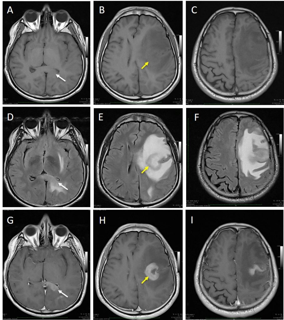
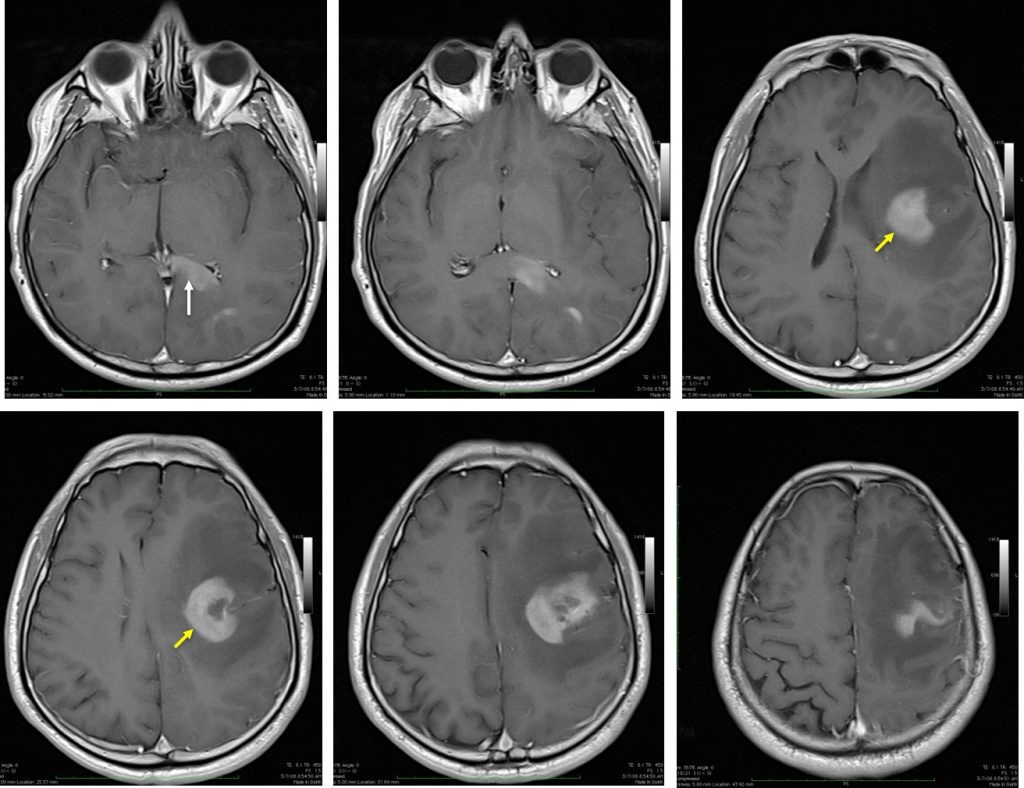
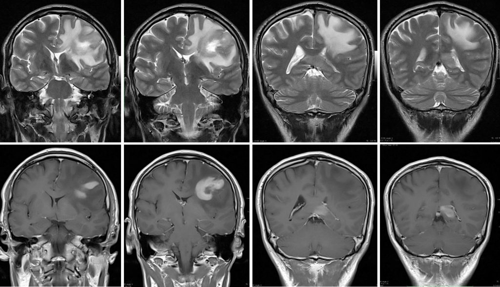
- There is a lesion at the left frontal region with marked surrounding vasogenic oedema (yellow arrows).
- It enhances post contrast, with evidence of central necrosis .
- There is mass effect onto the body of the ipsilateral lateral ventricle with slight midline shift to the contralateral side.
- Similar enhancing lesions with significant vasogenic oedema are seen in the left posterior parietal lobe and also splenium of the corpus callosum (white arrows).
- No evidence of hydrocephalus.
- There is effacement of the left cerebral sulci.
Progress of patient:
- Based on MRI findings, with presence of central necrosis GBM was given as diagnosis.
- Left frontotemporal craniotomy and tumour excision was done.
- HPE came back as B-cell lymphoma
- CT scan of neck, thorax, abdomen and pelvis show no significant finding.
- Patient was started on chemotherapy and shows good respond.
- After 4 years remission, patient presented again with short term memory loss and abnormal behaviour
- Repeat MRI shows more typical appearance of CNS lymphomas with multifocal periventricular lesion suggestive of recurrence.
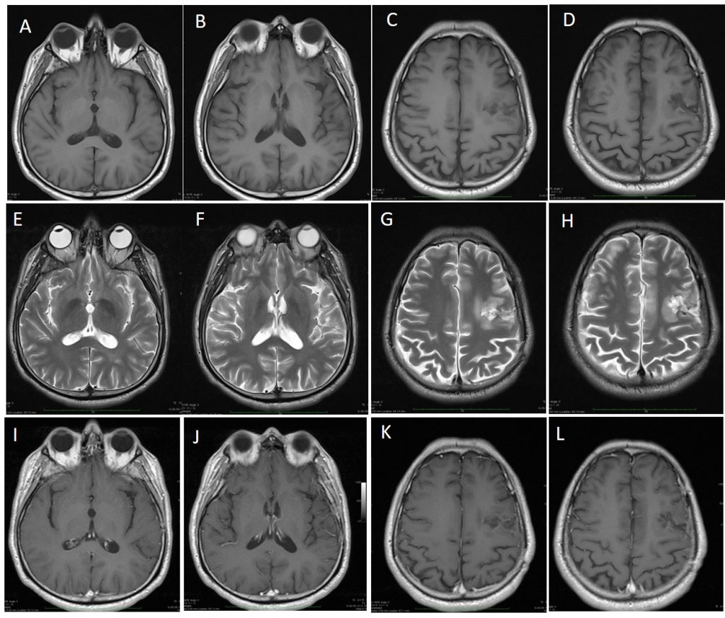
Final diagnosis: Primary CNS lymphoma with recurrence
Discussion:
- Primary CNS lymphoma are previously rare, currently relatively more commonly seen, ranks behind meningioma and low grade gliomas.
- It accounts for 2.5% of all brain tumours.
- Typically patients are over the age of 50 with a short duration of symptoms.
- There is a male predominance of approximately 2:1
- Classic imaging appearance for primary CNS lymphoma is of a CT hyperdense avidly enhancing mass, with T1 hypointense, T2 iso- to hypointense, vivid homogeneous gadolinium-enhancing lesion(s) with restricted diffusion on MRI, and exhibiting subependymal extension and crossing of the corpus callosum.
Recent Comments