
Clinical:
- An 80 years old lady
- Underlying DM and HPT
- Patient admitted for osteoporotic compression fracture of L1
- Also complains of abdominal pain and constipation
- No PR bleed
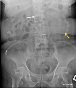
Radiographic findings:
- A rounded opacity seen at left lumbar region (yellow arrow).
- It is well defined and seen overlying the lower part of left renal shadow.
- There is no calcification seen within the lesion.
- There is no obliteration of the left psoas margin.
- Reduction in height of L1 vertebra is seen (white arrow)
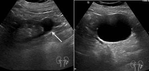
Ultrasound findings:
- Both kidneys have normal parenchymal echogenicity and size.
- No hydronephrosis or renal calculus bilaterally.
- There are multiple exophytic cysts at the lower pole of left kidney. The largest measuring 6.3 x 6.5 x 6.5cm (AP x W x CC). These cystic lesions have no internal septations, peripheral calcification or echogenic debris within.
- No free fluid within the abdomen.
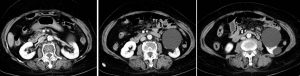
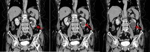
CT scan findings:
- Both kidneys are normally located.
- A few non enhancing exophytic cysts are seen in both kidneys.
- The largest exophytic cyst (mean HU: 4) is seen in the left lower pole measuring about 6.4 (AP) x 6.4 (W) x 7.4cm (CC).
- No internal septation, wall calcification or solid component seen.
Diagnosis: Simple renal cysts (Bosniak I)
Discussion (Bosniak classification):
- Bosniak I: Simple cyst with imperceptible wall, no risk of malignancy, no need work-up
- Bosniak II: minimally complex cyst with thin septa (<1mm) or thin calcifications. Can have hemorrhagic component. Renal lesion <3 cm and well marginated. No need work-up. No risk of malignancy
- Bosniak IIF: minimally complex cyst with increased number of septa or minimally thickened with nodular or thick calcifications. Minimal thickening of the wall with perceivable enhancement. If hyperdense cyst >3 cm mostly intrarenal and less than 25% of wall visible with no enhancement. This lesion need follow up at reasonable 6 months period. Percentage of malignancy in 5%.
- Bosniak III: indeterminate. Cyst with thick, nodular multiple septa or wall with measurable enhancement. Needs partial nephrectomy or radiofrequency ablation. Percentage malignant is 55%.
- Bosniak IV: clearly malignant. A solid mass with a large cystic or a necrotic component. Partial or total nephrectomy recommended. Percent of malignancy: 100%.
Recent Comments