
Clinical:
- A 48 years old lady
- Underlying DM and HPT
- Presented with left flank pain for 2 weeks
- Associated with fever
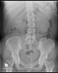
Radiographic findings:
- Prominent soft tissue shadow at left lumbar region.
- Minimal indentation to medial wall of bowel loops.
- No calcification seen. Bones are unremarkable.
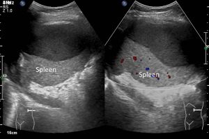
Ultrasound findings:
- A well-defined homogenous subcapsular splenic collection with layering sediments is seen compressing the splenic parenchyma, measuring about 10.7cm x 5.0cm.
- No vascularity or calcification within.
- Otherwise the spleen is normal in size and splenic parenchyma appears homogenous.
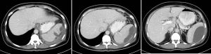
CT scan findings:
- The spleen is enlarged. Presence of a large subcapsular collection mainly laterally measuring about 10.3cm (AP) x 4.3cm (W) x 8.6cm (CC).
- There is associated fat streakiness adjacent to the collection.
- No septation, calcification or solid component is seen within the collections.
- The left hemi-diaphragm appears elevated and thickened with adjacent minimal left pleural effusion seen.
- Presence of left pleural effusion and basal atelectasis on the left side.
Diagnosis: Splenic abscess
Discussion:
- Splenic abscesses are uncommon
- The main causes include immunodeficiency conditions, hematogenous spread of distant infection, contiguous infection from adjacent infection such as perinephric abscess, trauma or from splenic infarction.
- Ultrasound appearance ranges from predominantly hypoechoic to hyperechoic with internal echoes. They may contain septa of varying thickness.
- CT scan normally shows low density (HU20-40) with minimal peripheral enhancement. Ascites and adjacent pleural effusion is commonly seen.
Progress of patient:
- Percutaneous drainage of abscess done
- Patient treated with antibiotic
- HIV, Hepatitis screening negative
- Connective tissue screening is also negative
- Cytology report of splenic aspirate consistent with abcess. No suspicious cell seen.
- Culture of splenic aspirate grows E.coli.
- AFB negative
Recent Comments