
Clinical:
- A 21 years old lady
- Previously healthy
- Presented with low back pain, right lower limb pain, shooting upward for few months.
- Later also developed numbness of right lower limb.
- Normal neurological assessment.
MRI findings:
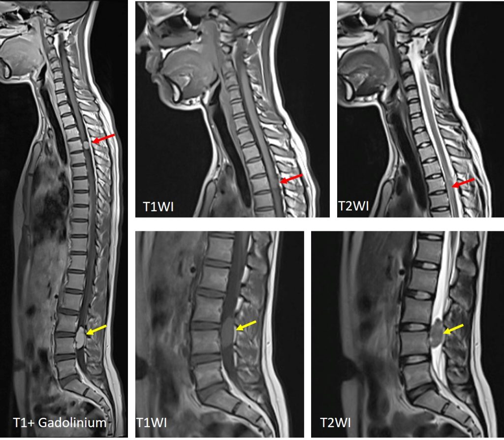
- There are two lesions seen within the spinal canal.
- The upper lesion located at T3 level is isointense on T1 and slightly hyperintense on T2WI compared to the cord
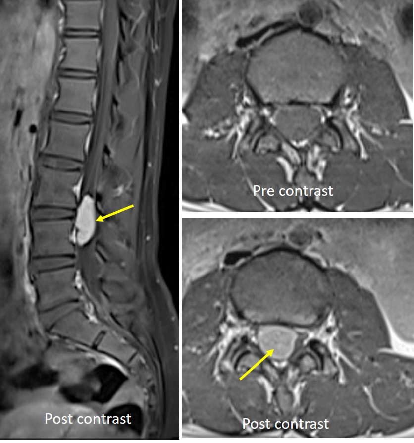
- The lower lesion at L3-L4 levels is isointense on T1 and hypointense on T2WI compared to the cord.
- Both lesions show marked enhancement post contrast
- The lesions are intradural and extramedullary
- No expansion of the neural foramina.
- Compression and distortion of the spinal cord is seen. No abnormal signal intensity within the cord itself.
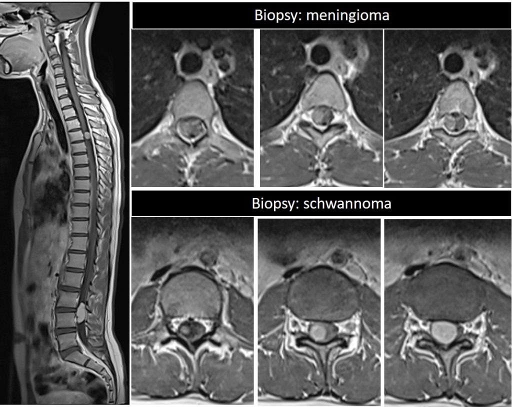
HPE findings: Upper lesion (Psammomatous meningioma, WHO Grade 1) and lower lesion (schwannoma).
Discussion:
- Schwannoma and meningioma are common differential diagnoses for intradural extramedullary spinal tumours. Other include neurofibroma and ependymoma.
- Half of cervical tumours were schwannomas, 72% thoracic lesions were meningiomas and all lumbar tumours are schwannomas
- Meningiomas were significantly located at the upper and mid-thoracic levels and schwannomas in lumbar area
- T1WI; no statistical difference. T2WI; schwannomas hyperintense and heterogenous.
- Post contrast: Schwannomas intense and heterogenous, Meningioma : moderate and homogenous.
- Meningioma: Dural tail sign, bone sclerosis.
- Schwannoma: neural foraminal extension and foraminal widening
Recent Comments