
Clinical:
- A 3 years old boy with bilateral foot deformity noted since birth.
- Late on walking. Also has rotated left leg.
- There is associated bowel symptoms; on & off constipation and diarrhoea.
- Neurologically, unable to assess fully.
- MRI TRO spinal cord deformity.
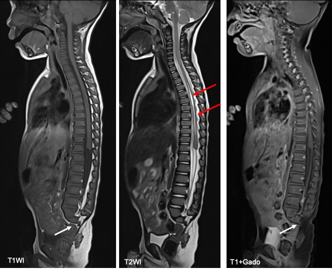
MRI findings:
- The cervical, thoracic and lumbar spine are normal.
- There are only S1 and portion of S2 present. The rest of the sacrum and coccyx are absent (white arrows).
- The conus medullaris lies at T12.
- The spinal canal up to its termination level is not narrowed.
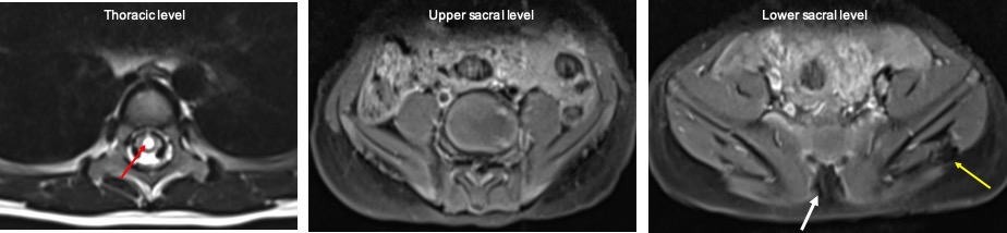
- There is a tubular structure within the thoracic spinal cord extending from the T3 to T12 which shows high signal intensity on T2 & low on T1 in keeping with syrinx (red arrows).
- The gluteus muscles are atrophied; more on the left side (yellow arrow) with increased adipose tissue at the gluteus region. Some fatty replacement is also noted within the gluteus muscles.
- No abnormal meningeal outpouching seen. No communication externally.
- No focal extramedullary or epidural lesions seen within the spinal canal.
Radiological diagnosis: Sacral agenesis with syringomyelia.
Discussion:
- Sacral agenesis is considered a part of caudal regression syndrome.
- It is a rare sacral developmental abnormality consisting of absence of part or all of the sacrum.
- Sacral agenesis has an incidence of 1 in 25,000 live births and is associated with maternal diabetes in approximately 25% cases.
- It is clinically associated with malformation of the hindgut, caudal spinal cord, lower limbs, and the urogenital system.
- Multiple vertebral anomalies such as fused vertebra, hemivertebra, butterfly vertebra, and diastematomyelia may also be seen.
- Renshaw classified sacral agenesis into four types, based on the amount of sacrum remaining and articulation between pelvis and spine.
- type I:unilateral agenesis localized to sacrum or coccyx
- type II:partial agenesis with bilateral defects; the iliac bone articulates with S1, but the distal sacral elements fail to develop
- type III:total sacral agenesis; iliac bones articulate with the lowest lumbar element
- type IV:total sacral agenesis; iliac bones fuse posteriorly
Recent Comments