
Clinical:
- A 30 years old lady
- Investigation for infertility
- Ultrasound shows retroverted uterus
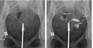
Hysterosalpingogram findings:
- Uterus in retroverted.
- Uterine cavity shows normal outline and no filling defect within it
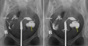
- There is spillage from right side (white arrows).
- The proximal left fallopian tube is normal in calibre.
- However the tube is dilated distally with no evidence of contrast spillage on the left side (yellow arrows)
Radiological diagnosis: Left hydrosalpinx
Discussion:
- A hydrosalpinx is a condition that occurs when the fallopian tube is blocked and fills with fluid.
- If the fluid is infected, i.e. pus, then it is a pyosalpinx and if it is bloody, it is then haematosalpinx.
- The blocked tube may become substantially distended giving the tube a characteristic sausage-like or retort-like shape.
- The condition is often bilateral and the affected tubes may reach several centimeters in diameter. The blocked tubes cause infertility.
- Hydrosalpinx may be diagnosed using ultrasoundas the fluid filled elongated and distended tubes display their typical echolucent pattern. However, a small hydrosalpinx may be missed by sonography.
- Hysterosalpingogram )HSG) shows the retort-like shape of the distended tubes and the absence of spillage of the dye into the peritoneum. If, however, there is a tubal occlusion at the utero-tubal junction, a hydrosalpinx may go undetected.
- MRI and CT scan may also demonstrate the dilated fallopian tubes.
Recent Comments