
Case contribution: Prof. Ahmad Sobri Muda
Clinical:
- A 35 years old lady who is a housewife
- Complaint of low back pain for the past few years which recently worsening.
- Currently she had the pain almost every night and has to sit when praying. She has to change position frequently during sitting and standing to relieve the back pain.Her VAS is 5-6/10.
- Investigations show evidence of degenerative disc disease with mild facet artropathy.
- Further clinical evaluations indicate that the pain is arising from facet joints rather than pure discogenic pain.
- Facet joint injection with combination of steroid and anesthetic drugs performed.
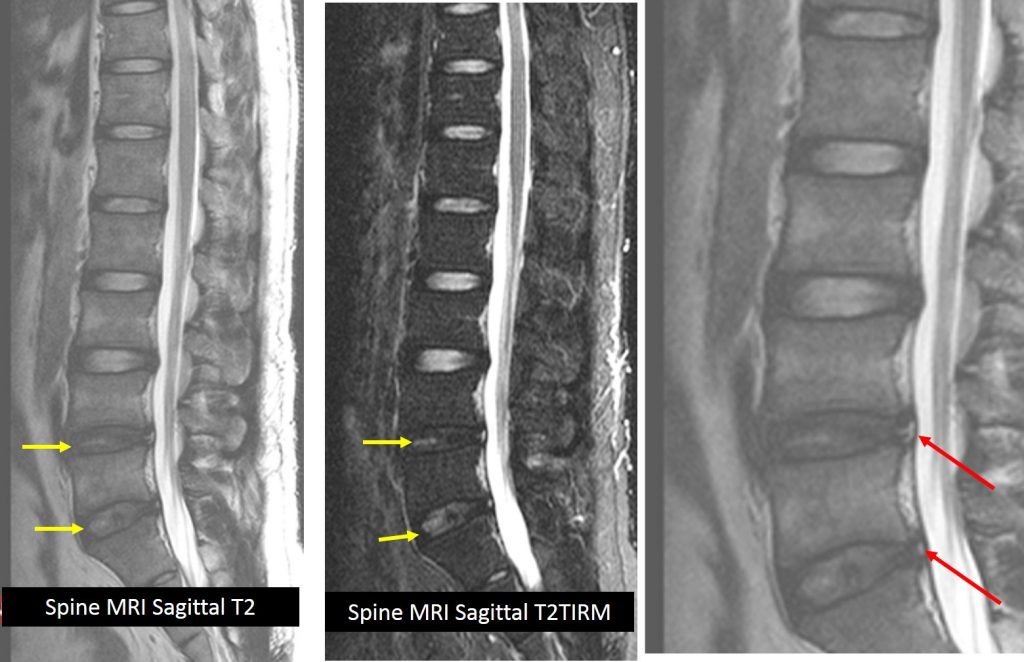
MRI findings:
- MRI shows degenerative disc changes most prominent in L4/L5 and L5/S1 (yellow arrows).
- There are high intensity zone (HIZ) within the posterior annular of L4/L5 and L5/S1, (red arrows) suggestive of posterior annular tear.
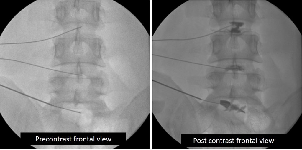
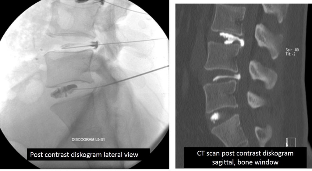
Diskogram findings:
- Lateral view of diskogram showing contrast leak beyond central outline of nucleus pulposus, indicating posterior annular tear at the level of L3/L4 and L4/L5.
- Sagittal reconstruction of CT Scan post diskogram confirms the contrast leak at posterior annular tear of L3/L4 and L4/L5.
Diagnosis: Posterior annular tear
Discussion:
- Annular tears or annular fissures are degenerative deficiency of annulus fibrosus of the intervertebral disc.
- Most are asymptomatic, however, some are painful. The defect allows in-growth of nerve endings and granulation tissue.
- Fissures near the dorsal root ganglion are especially likely to be painful.
- Annular tears of the intervertebral disc can occur due to a many causes. Commonly they do occur as our discs naturally degenerate with age. Other factors contributing to annular tears include certain occupations or high impact activities putting intervertebral discs under added pressure.
- Although very common, only a minority are identified on MRI examination
- It is characterised by a region of high T2 signal (high intensity zone) in the otherwise low signal annulus.
- Most annular tears improve and heal over time with rest, physical therapy and anti-inflammatories medication.
- In some situations symptoms may warrant some intervention.
Progress of patient:
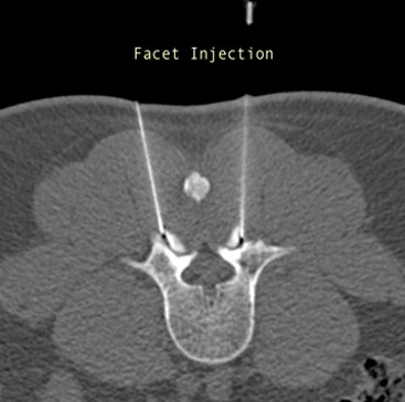
- Clinically patient’s presentations are in from the facet atropathy, thus intervention performed to alleviate the pain.
- Facet injection with mixture of steroid and local anesthesia performed under CT guidance.
- Patient’s sign and symptoms improved significantly with the facet injections
Acknowledgement:
- Dr. Roger Tok Chung Hong
- Dr. Naveen Rajadurai
- Dr Muhammad Fadli Embong
References/Suggested reading:
- Image-guided facet joint injection. Peh WCH. Biomed Imaging Interv J 2011; 7(1)
- Lumbar disc nomenclature; version 2.0. Recommendations of the combined task force of the North American Spine Society, the American Society of Spine Radiology and the American Society of Spine Radiology, Fardon et al. The Spine Journal 14 (2014): 2525-2545.
- Progression of lumbar disc herniations over an eight-year period in a group of adult Danes from the general population – a longitudinal MRI study using quantitative measures. Kjaer et al. BMC Musculoskeletal Disorders (2016) 17:26.
Recent Comments