
Case contribution: Dr Radhiana Hassan
Clinical:
- A 20 years old man
- No known medical illness
- Presented with right scrotal swelling for one month
- No fever, non-tender
- No constitutional symptoms
- AFP=94.6 IU/mL (normal <7.4)
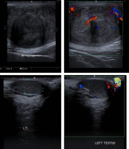
Ultrasound findings:
- There is a large testicular mass measuring about 7.0 x 5.0 cm
- It shows heterogeneous hypoechogenicity with no obvious cystic component or calcification within it
- On Doppler, there is increased colour flow in the periphery of mass
- The scrotal wall is thickened and heterogenous.
- The left testis is pushed superoposteriorly in the left scrotum. It is normal in size measuring 3.1 x 1.2 cm. It shows homogenous echogenicity with no focal mass lesion within.
Diagnosis: Testicular cancer (HPE: embryonal cell carcinoma)
Discussion:
- Testicular cancers are the most common neoplasm in men between the ages of 20 and 34 years.
- Risk factors: cryptochidism, family history, radiation, previous contralateral testicular tumour, microlithiasis, hypospadia, infection, infertility and Klinefelter syndrome
- More than 90% are primary tumours and about 90% are testicular germ cell tumour
- Secondary tumour include secondary testicular lymphoma (most common testicular malignancy in older men), testicular leukaemia and metastasis to testis
- Testicular embryonal cell carcinoma is a type of non-seminomatous germ cell tumour with peak incidence at around 25-30 years of age.
Progress of patient:
- Orchidectomy done
- Planned for chemotherapy after operation
- Patient request postponed chemotherapy to finish his study
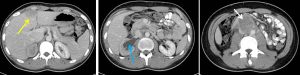
- 4 months later, repeat CT scan show metastasis to lung, liver and aortocaval nodes

Recent Comments