
Case contribution: Dr Radhiana Hassan
Clinical:
- A 63 years old lady
- Underlying DM on treatment
- Presented with sudden onset of severe, dull aching abdominal pain at right side
- No aggravating or relieving factor
- Associated with vomiting 4-5 times since having pain
- No loose stool, no altered bowel habit, no PR bleed
- BP=128/84 mmHg, HR= 88 bpm, T=36.8
- Pain score 7/10
- Clinically there is tenderness and guarding at right iliac fossa with vague mass
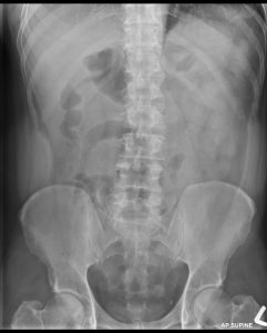
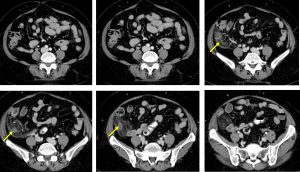
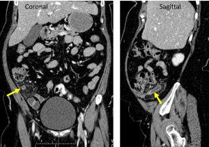
CT scan findings:
- Transverse retrocecal appendix is noted (yellow arrows).
- Appendix is dilated measuring up to 1.2 cm in diameter. Its wall appears thickened and irregular.
- The surrounding fat is streaky. No obvious collection at right iliac fossa region.
- No pneumoperitoneum. No free fluid.
- The rest of bowel loop is neither thickened nor dilated. No pelvic free fluid.
- Uterus is normal. No adnexal lesion is seen.
Intra-operative findings:
- Laparoscopic appendicectomy done
- Suppurative appendicitis
- Minimal hemoserous fluid at right iliac fossa and pelvis
- No pus. Base of caecum healthy
- HPE: acute appendicitis
Diagnosis: Acute appendicitis
Discussion:
- Transverse retrocaecal appendix comprise about 2% of all appendix location.
- CT is highly sensitive (94-98%) and specific (up to 97%) for the diagnosis of acute appendicitis
- CT findings include:
- appendiceal dilatation (>6 mm diameter)
- wall thickening (>3 mm) and enhancement
- thickening of the cecal apex
- periappendiceal inflammation
- focal wall nonenhancement representing necrosis (gangrenous appendicitis) and a precursor to perforation
- appendicolith
- periappendiceal reactive nodal enlargement
Reference:
- https://radiopaedia.org/articles/appendicitis-2
Acknowledgement:
- Dr Siti Kamariah Che Mohamed
Recent Comments