
Case contribution: Dr. Radhiana Hassan
Clinical:
- A 62 years old lady
- Underlying DM, HPT and hyperlipidaemia
- Also had gallstone, refused operation due to financial constraint
- Presented with right hypochondriac pain, fever and reduced oral intake
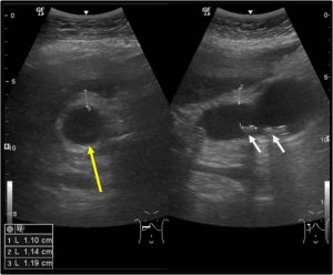
Ultrasound findings:
- Gallbladder is distended with multiple echogenic foci casting acoustic shadowing is seen within it in keeping with calculi (white arrows).
- The largest calculus measures about 1.1 cm in length.
- No sludge or sediment is seen within the gallbladder.
- The gallbladder wall appears thickened measuring up to 1.2 cm in maximum thickness.
- Presence of pericholecystic fluid is also observed.
- Sonographic Murphy’s sign is positive.
- The biliary tree is not dilated with CBD measures 3.9 mm in diameter.
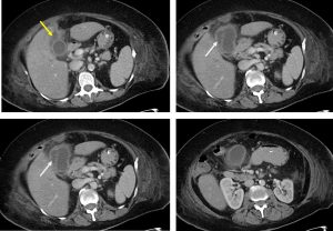
CT scan findings:
- The gallbladder is grossly distended and filled with homogenous fluid, which is slightly denser at the dependant part (HU average: 20-30).
- The gallbladder wall is thickened and oedematous measuring up to 1.0 cm in thickness.
- No luminal or intra-mural air pocket is detected.
- There is adjacent fluid collection which is seen tracking superiorly under the liver and anteriorly to the abdominal wall.
- Pericholecystic fluid and the surrounding fat streakiness is also observed.
- Minimal interloop fluid is also seen inferior to the liver and below the spleen.
- Significant free fluid is also noted at the pelvic region.
Progress of patient:
- Patient conditioned worsened due to sepsis.
- Patient also developed respiratory distress requiring intubation and admission into ICU after one day admitted.
- Percutaneous cholecystostomy done by IR and about 10 cc of pus aspirated.
- Pus C&S from cholecystostomy: Klebsiella pneumonia
- Completed antibiotic 10 days
- Complicated by Pulmonary embolism
- Discharged well later and planned for elective cholecystectomy.
Diagnosis: Gallbladder empyema
Discussion:
- Gallbladder empyema is an unusual complication of cholecystitis.
- Gallbladder lumen is distended and filled with purulent material (pus).
- Increased incidence in those with DM and/or advanced atherosclerotic disease.
- Ultrasound may show the usual sonographic features of cholecystitis with added echogenic content within the gallbladder lumen (not specific for an empyema).
- CT may show general imaging features of cholecystitis with added high-attenuating material (representing pus) within the distended gallbladder lumen. Again this feature is nonspecific and is often difficult to differentiate from sludge within the gallbladder.
Recent Comments