
Image 1:
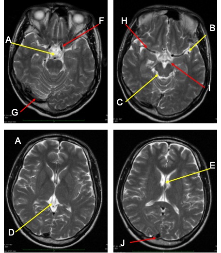
- Name the examination
- Name the labelled cisterns/CSF space/ventricles (yellow arrow: A-E)
- Name the vessels (red arrows: F-J)
Image 2:
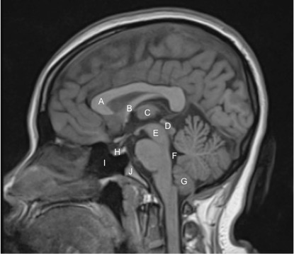
- Name the examination
- Name the labelled structures
Image 3:
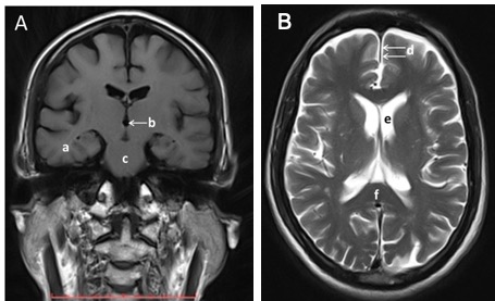
- Name the examinations
- Name the labelled structures.
Answers:
Image 1:
- MRI brain in axial planes, T2-weighted image
- The labelled structures:
- Interpedicular cistern
- Left sylvian fissure
- Right ambient cistern
- Quadrigeminal cistern
- Left frontal horn of lateral ventricle
- Left Internal carotid artery
- Left sigmoid sinus
- Left middle cerebral artery
- Left posterior cerebral artery
- Transverse sinus
Image 2:
- Name of examination: MRI brain in sagittal plane, T1-weighted image
- The labelled structures:
- A. Genu of corpus callosum
- B.Fornix
- C.Massa intermedia/thalamus
- D. Quadrigerminal plate of midbrain
- E. Midbrain
- F. Fourth ventricle
- G. Cerebellar tonsil
- H. Pituitary gland
- I. Sphenoid sinus
- J. clivus
Image 3:
- The examinations:
- MRI of brain, coronal plane, T1 weighted image, non-contrast
- MRI brain, axial plane, T2-weighted image
- The labelled structures:
- a) Right temporal lobe
- b) third ventricle
- c) pons
- d) anterior falx cerebri
- e) frontal horn of left lateral ventricle
- f) corpus callosum
Recent Comments