
Case contribution: Dr Radhiana Hassan
Clinical:
- A 70 years old man
- Involved in motor vehicle accident
- Underlying DM, CKD stage III and CVA with left hemiparesis
- On arrival at ED, BP=112/70mmHg, PR=100 bpm, GCS=15/15
- Non-contrast CT abdomen requested to rule out intra-abdominal injury
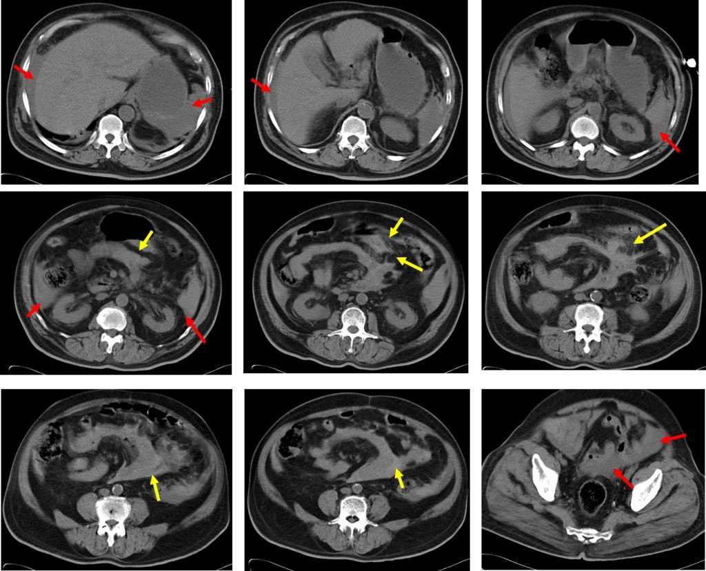
CT scan findings:
- There is massive hemoperitoneum with blood collections seen at perihepatic, perisplenic, both pericolic gutter and pelvic region (red arrows)
- There is blood collection at inter-mesenteric region with mesenteric fat streakiness (yellow arrows)
- No bowel wall dilatation or bowel wall thickening seen.
- No free intraperitoneal air
- Limited assessment of non-contrast study shows no obvious solid organ injury
Intra-operative findings:
- Mesenteric tear seen at distal third of transverse colon
- Bleeding seen from mesenteric vessels
- Transverse colon wall intact
- Estimated blood loss 2000 mls
- Rest of organ is normal.
Diagnosis: Mesenteric injury
Discussion:
- Mesenteric injury from blunt abdominal trauma is rare and can be difficult to diagnose
- Mesenteric fat infiltration or “stranding” can be associated with mesenteric injury with or without bowel perforation
- A localized hematoma within the mesentery in the absence of a bowel abnormality points to an isolated laceration of a mesenteric vessel
- Hemoperitoneum is a common finding in patients with intraperitoneal bowel or mesenteric laceration
- It follows that hemoperitoneum in the absence of solid organ injury would imply bowel or mesenteric laceration as the source of bleeding
- Definitive CT signs include active contrast extravasation of contrast media, intermesenteric free fluid forming triangles, beading and termination of mesenteric vessels, abrupt termination of vessels and also pooling of contrast on multiphase scan
Recent Comments