
Case contribution: Dr Radhiana Hassan
Clinical:
- A 34 years old lady
- No known medical illness
- Complaint of swelling at ankle region for one year, recently associated with pain
- Clinical examination shows a soft swelling at left lateral malleolus region
- No overlying skin changes. Slight reduction in range of motion.
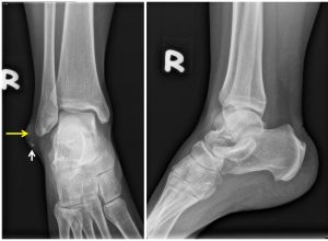
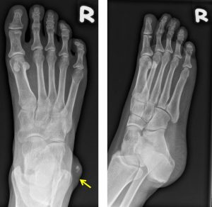
Radiographic findings:
- There is soft tissue swelling at lateral malleolus region (yellow arrows)
- No thickening of overlying skin. No skin break seen.
- A few calcifications within the soft tissue swelling (white arrow)
- No sclerotic or lytic changes of the adjacent bone. No periosteal reaction.
Progress of patient:
- Excision done under LA
- Patient recovered well
HPE findings:
- Macroscopy: specimen labelled as right ankle lump consists of a piece of skin tissue measuring 25x14x12 mm. Cut section shows a uniloculated cyst in the dermis measuring 10x6x3 mm. The cyst contain sebaceous material with cyst wall measures 1-2 mm thickness. No solid area seen.
- Microscopy: section shows a skin tissue composed of epidermis and dermis. The epidermis is unremarkable. There is a cystic lesion in the dermis partly lined by a keratinised stratified squamous epithelium and partly devoid of epithelial lining, replaced by chronic inflammatory cells. Scattered multinucleated giant cells are noted. The cavity cell is filled with lamellated keratin flakes. In areas, calcifications are seen. Negative for malignancy.
- Interpretation: Epidermal cyst.
Diagnosis: Epidermal cyst at right ankle
Discussion:
- It is lso known as epidermal inclusion cyst, sebaceous cyst (misnomer as the lesion do not originate from sebaceous gland) or epidermoid cyst
- It is the most common cyst of the skin
- The size ranges from a few milimeters to a few centimetres
- The contents are cheesy, malodorous mixture of degraded lipid and keratin.
- It can be found anywhere but typically seen on the scalp, face, neck, trunk and back.
- A fewer than 10% of cases occur in the extremities.
- Radiographic feature shows a well-circumscribed lesion arising in or just deep to the skin. Ultrasound it is predominantly hypoechoic and if small may mimic typical anechoic cyst. Larger lesion may be heterogenous due to presence of mucoid, fat, calcification or pus. Usually no associated vascularity seen. CT density is similar with water. On MRI it also shows signal intensity similar with CSF. Some shows restricted diffusion on MRI and some shows superimposed T2 shine through. No enhancement is seen on post contrast image.
- The differential diagnosis for unruptured cyst include ganglion or bursitis. Neurogenic tumours, nodular fasciitis, myxoid tumours and dermatofibrosarcoma shows variable enhancement on post contrast imaging.
- Ruptured cyst may be difficult to differentiate from abscesses or complicated ganglion or bursitis
- It rarely undergo malignant degeneration to squamous cell carcinoma.
- Other complications include superimposed infection, rupture and concurrent occurrence of other tumours .
Recent Comments