
Case contribution: Dr Radhiana Hassan
Clinical:
- A 63 years old lady
- Had underlying hypertension and CVA with left sided hemiparesis, ADL semi dependent walking with walking stick at home
- Referred from GP
- Patient noted right breast lump on SBE
- Gradually increasing in size, non tender
- No nipple discharge, no skin changes, no LOA, no LOW
- No family history of breast cancer
- Breastfed all 5 children up to 5 years
- Clinically a palpable mass at right axillary tail measuring 3×3 cm, hard, attached to the skin not to the chest wall, peau de orange +ve. No nipple retraction, right axillary node palpable.
- Left breast is normal, left axilla is also normal
Mammogram findings:
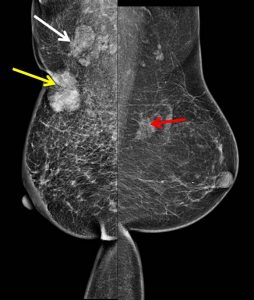
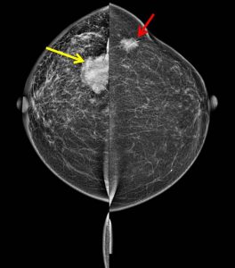
- There is an ill-defined oval high density lesion observed in the upper outer quadrant of the right breast (yellow arrows) with associated stromal distortion and skin traction.
- No clustered microcalcification seen within the lesion.
- A few lobulated lesions are seen at right axillary tail region.
- An ill-defined lesion with associated stromal distortion is also seen in the upper outer quadrant of the left breast. No calcification is seen within this lesion.
- Skin thickening is seen at right periareolar region.
- No nipple retraction bilaterally.
Ultrasound shows suspicious lesions in both breasts (images not shown)
HPE finding:
- Invasive carcinoma of right breast lesion
- Invasive carcinoma of left breast lesion
Diagnosis: Bilateral synchronous breast cancer
Discussion:
- Synchronous breast cancers are two or more primary breast cancers that occur in either breast at the same time
- About 1.4-12% of all breast cancers may be synchronous
- Bilaterality is greatest with invasive lobular carcinoma
- It is important to consider metastasis to the breast from opposite breast (which is unusual especially with no other evidence of metastasis)
- It has worse survival rates because of distant metastasis
Progress of patient:
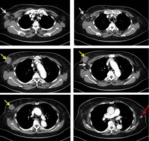
- CT scan for staging do not demonstrate distant metastasis
- The right and left breast lesion with lymphadenopathies are again seen
- Patient had chemotherapy at another hospital and lost to follow up
Recent Comments