
Case contribution: Dr Radhiana Hassan
Clinical:
- A 61 years old lady
- Presented with chronic headache for 4 years
- Associated with giddiness and blurred vision
- On and off fever, no body weakness
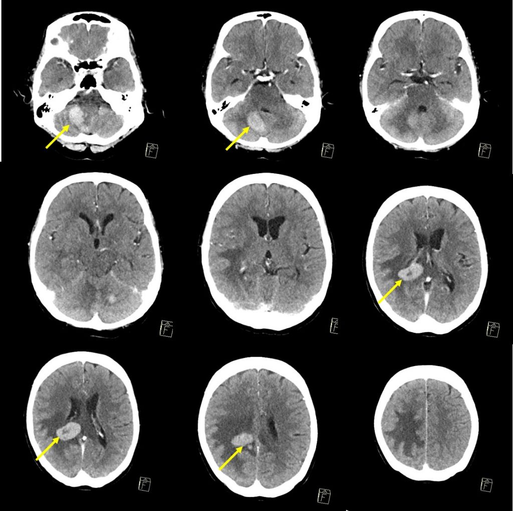
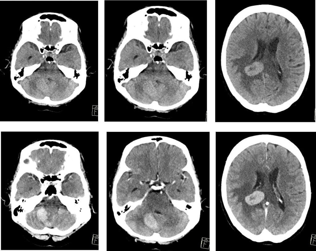
CT scan findings:
- Hyperdense lesions are seen at white matter of right parietal and right cerebellum
- The lesions are located at periventricular region
- It shows marked enhancement post contrast
- Areas of central non-enhancing hypodensity is seen
- Significant perilesional oedema is seen
- No midline shift or hydrocephalus
Diagnosis: Primary CNS lymphoma (HPE proven)
Progress of patient:
- CT scan thorax, abdominal pelvis are normal
- Burrhole biopsy done, HPE revealed DLBCL
- Completed chemotherapy, shows good response
- Planned for radiotherapy
Discussion:
- Primary CNS lymphoma is an uncommon brain tumours.
- On imaging, characteristically is is seen as hyperdense enhancing supratentorial lesion as in this case.
- However, usually the enhancement is intense with no area of necrosis.
- There is little associated vasogenic oedema.
- Mainly the lesions are solitary (60-70%); multiple lesion seen in 30-40%
- There is predilection for periventricular white matter.
- But it can also occur in cortex or deep gray matter
- It is most frequently found in supratentorial brain (70%)
Recent Comments