
Case contribution: Dr Radhiana Hassan
Clinical:
- A 0ne year old girl
- First twin of monochorionic diamniotic twin
- She has dysmorphysism with retrognathia, microcephaly and global developmental delay
- Also has reducible umbilical hernia suckling and swallowing incoordination
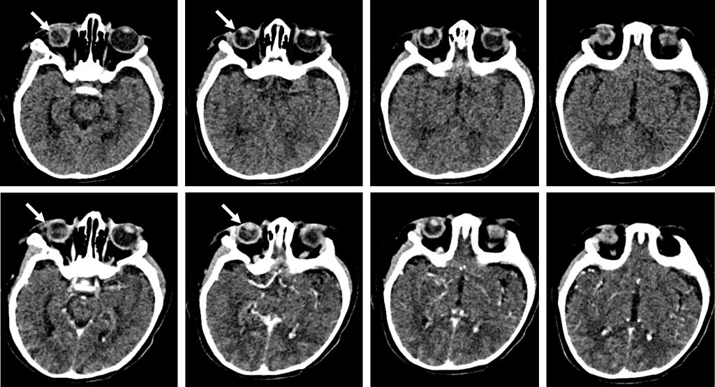
CT scan findings:
- The right globe is smaller than the left side
- Soft tissue density lesion within the right globe
- Slight increase in density of right vitreous
- There is no obvious calcification within it
- No contrast enhancement seen on post contrast
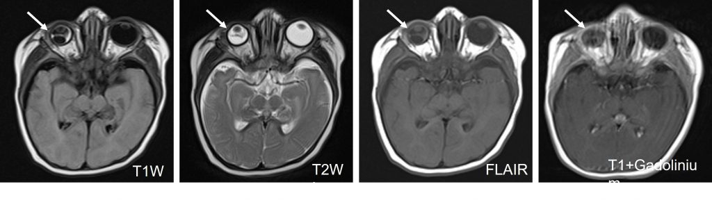
MRI findings:
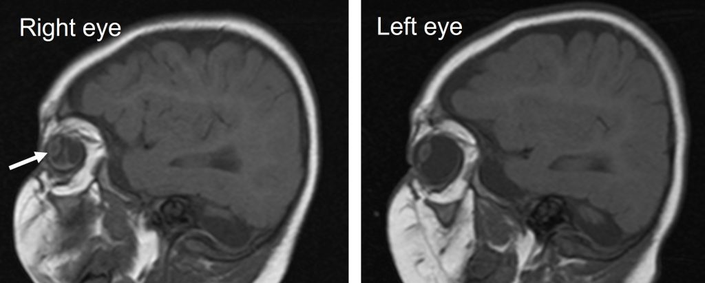
- The right globe is smaller
- A posteriorly detached soft tissue with fluid noted posterior to it
- No area of enhancement seen
- Extra ocular muscles and optic nerves are normal bilaterally
- Dandy walker malformation also noted (images not shown)
Diagnosis: Coats disease of the right eye
Discussion:
- Coats disease is a rare congenital disease affecting the eyes and is a cause of leukocoria
- It is also known as exudative retinitis or retinal telangiectasia
- It occurs predominantly in young patients with the vast majority are diagnosed before the age of 20 years
- There is well recognized male predominance (69-85%)
- The exact etiology is unknown and the disease is a non-hereditary disorder.
- The disorder is primarily one of weak capillaries of the retina, resulting in progressive retinal detachment due to an exudative subretinal collection.
- Clinical presentation is usually with leukocoria or strabismus, although eventually, complete blindness may develop.
- The diagnosis is usually made with indirect ophthalmoscopy.
- The disease is unilateral in 80-90% of cases.
- Ultrasound is an excellent modality for the assessment of globe, however has limited specificity and difficulty in distinguishing Coats disease from non-calcified retinoblastoma.
- CT findings depends on the stage of the disease. Enhancement may be seen at the margins of the exudate and may have a V-shaped pattern similar to retinal detachment. In advanced cases the affected globe is hyperdense due to proteinaceous exudates and the vitreous space may be obliterated due to extensive retinal detachment.
- Calcification is uncommon but has been reported.
- The affected eye in Coats disease is usually significantly smaller and is thought to represent an impairment of growth rather than the reduction in the volume of a previously normal globe.
- MRI appearances are similar to CT with better contrast resolution and the ability to visualize fainter enhancement, but reduced ability to visualize calcium. MRI is also able to visualize globe size.
- T1:high signal due to proteinaceous nature of the exudates
- T2:high signal due to proteinaceous nature of the exudates
- T1 C+ (Gd)-enhancement of the detached retina may be visible
Recent Comments