
Case contribution: Dr Radhiana Hassan
Clinical:
- A 30 years old lady
- Primary infertility for 7 years
- Menses regular and normal
- Transvaginal ultrasound was normal
- Hormonal essay is normal
- HSG to check tubal patency
Hysterosalpingography findings:
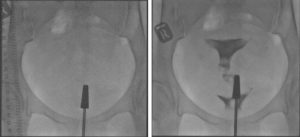
- Unremarkable preliminary image.
- Post contrast: Uterus and both Fallopian tubes are demonstrated.
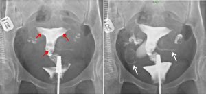
- The uterine cavity has smooth outline and normal in appearance
- Non-persistent filling defects within uterine cavity (red arrows) are likely to represent air bubbles.
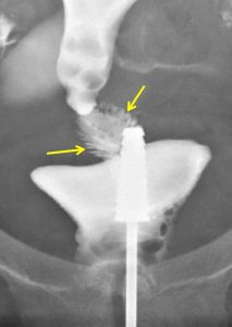
- Fine and numerous feathery-like appearances are seen at cervical canal (yellow arrows) in keeping with endocervical plicae palmatae.
- Both Fallopian tubes are normal and not dilated.
- Free peritoneal spillage is demonstrated from both Fallopian tubes (white arrows).
- Impression: Patent bilateral fallopian tubes.
Discussion: Cervical plicae palmatae
- Cervical plicae palmatae are normal folds seen on the anterior and posterior walls of the cervical canal.
- Studies report it in 45-50% of women between the ages of 20-50 years. It is most frequently seen in the fourth decade women.
- They are thought to be a remnant of mullerian duct fusion during fetal development.
- On HSG it is described as serrated appearance at endocervical region. They are often described as longitudinal ridges or oblique elevation.
- It can also be identified on ultrasound and MRI imaging.
- It should not be diagnosed as pathological findings.
Recent Comments