
Case contribution: Dr Radhiana Hassan
Clinical:
- A 49 years old man
- No known medical illness
- Presented with headache and fitting episode
- Developed one episode of status epilepticus
- No fever, no meniningism, muscle weakness involving left side of body
- Urgent non-contrast CT scan brain done was reported as cerebral infarction
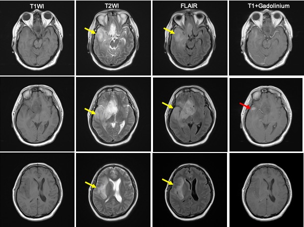
MRI findings:
- Abnormal signal seen involving right temporal, frontal and parietal lobe
- It is hypointense on T1, hyperintense on T2/FLAIR with minimal enhancement post contrast
- Cortex expansion is also noted
- Mass effect with midline shift and compression to ipsilateral lateral ventricles
- No restricted diffusion
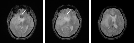
- Blooming artefacts within the lesion on hemo sequences
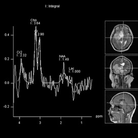
- MRS shows choline peak, decreased NAA
Diagnosis: Anaplastic astrocytoma (awaiting HPE result)
Discussion:
- Anaplastic astrocytoma is a diffusely infiltrating malignant astrocytoma
- It involves white matter with variable enhancement
- commonly involves the frontal and temporal lobe
- may involve and expand the cortex
- presence of flow voids or blooming artefact may be suggestive of progression to GBM
- Typically shows no restricted diffusion
- variable enhancement, but ring enhancement is suspicious of GBM
- MRS show elevatee Cho/Cr ratio, decreased NAA
Recent Comments