
Case contribution: Dr Radhiana Hassan
Clinical:
- A 48 years old male with underlying DM and HPT
- Admitted and treated for meningitis
- Noted to have abnormal sodium level during admission, correction done
- After 2 weeks patient requested AOR discharge from ward
- Presented again with fluctuant level of conciousness and spastic quadriparesis


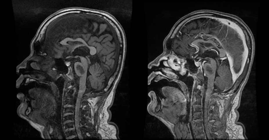
MRI findings:
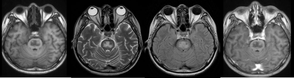
- A lesion seen within the pons, which is centrally located.
- It is hypointense on T1, hyperintense on T2/FLAIR, no restricted diffusion and not enhanced post contrast
- Sparing of peripheral and descending corticospinal tract region
- No swelling focal expansion of pons
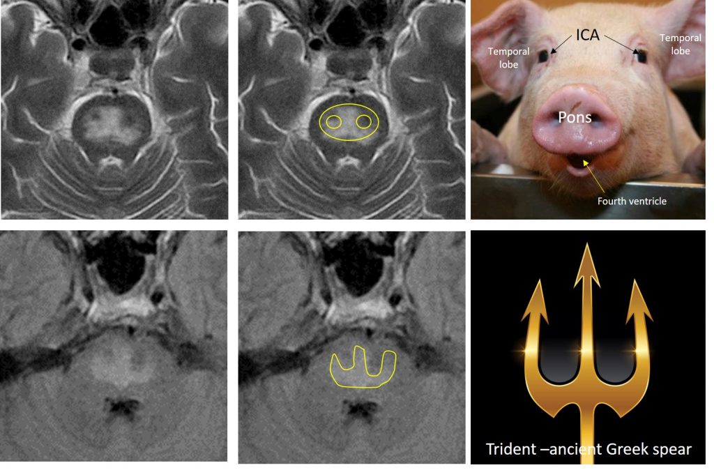
- Trident sign and piglet signs are seen
- A few deep white matter hyperintense foci on FLAIR sequence is seen.
- No periventricular or juxtacortical lesion
- No hydrocephalus, no abnormal leptomeningeal enhancement.
Diagnosis: Osmotic demyelination syndrome
Discussion:
- Osmotic demyelination syndrome refers to acute demyelination from rapid shift of osmolality, typically with the rapid correction of hyponatremia.
- It has replaced the term central pontine myelinolysis due to extra-pontine structures that can be affected in this condition.
- It is commonly seen in chronic alcoholics, chronically debilitated patients, transplant recipients and pregnancy-related hyperemesis.
- Imaging shows central pons T2-hyperintensity with sparing of periphery. Involvement is in pons (50%) and extrapontine in 50% (basal ganglia, cerebral white matter, hippocampi and central fibers). Regardless of site, demyelination often bilateral and symmetric
- Initial study may be normal, findings may be transitory, resolve completely, however persistent hyperintensity in 1-4 months can be seen (coagulative necrosis).
- On MRI classically it is mild/moderate hypointense on T1, hyperintense on T2/FLAIR in central pons with sparing of periphery, no hemorrhage, restricted diffusion in acute phase, usually does not enhanced.
- The piglet sign seen in osmotic demyelination syndrome. It refers to the appearance of upper pons in axial T2 or FLAIR. The abnormal T2-hyperintense signal within the pons reminiscent of a pig’s snout. The rest of the piglet features are formed by temporal lobes (ears), the carotid arteries (eyes) and the fourth ventricle (mouth)
- The trident sign describe the abnormal T2/FLAIR hyperintensity within the pons which is shaped like a trident (a three-pronged spear). The predominance involvement of the transverse pontine fibers and relative sparing of the descending corticospinal tracts is responsible for this characteristic appearance.
Recent Comments