
Case contribution: Dr Radhiana Hassan
Clinical:
- A 42 years old lady
- Came for screening mammogram
- No family history of breast cancer
Mammogram findings:
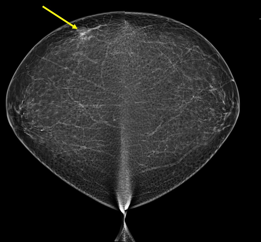
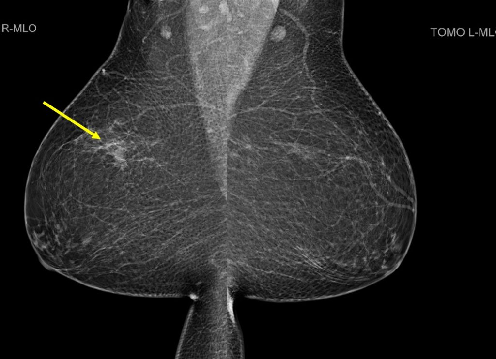
- The breasts are almost entirely fatty
- There is a focal density at right upper outer quadrant
- No obvious border to suggest mass lesion
- No obvious stromal distortion
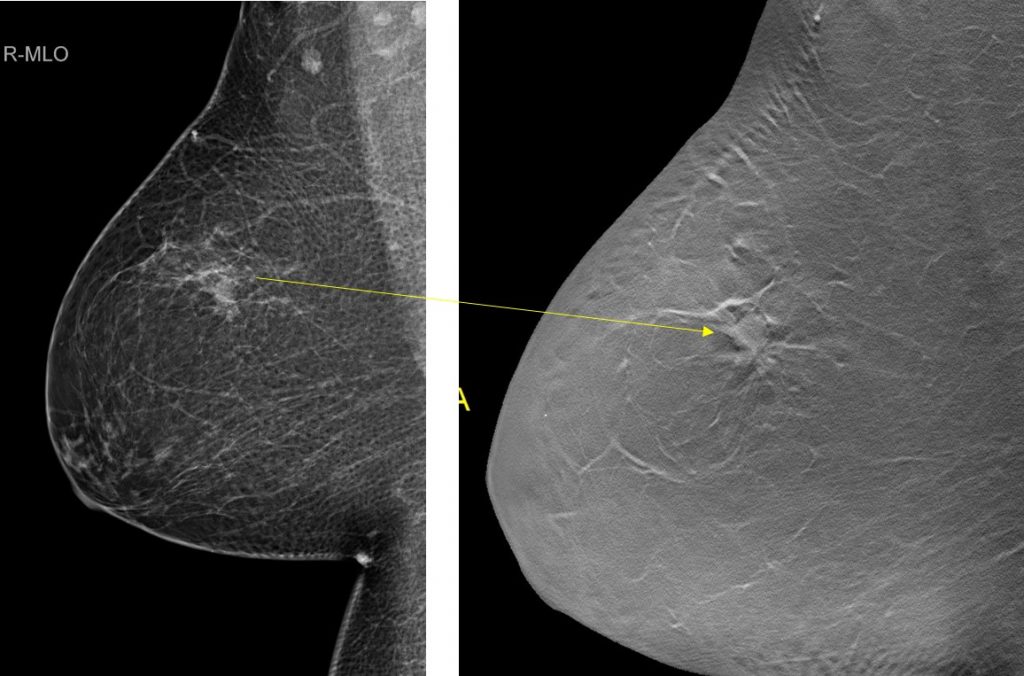
- However, on tomosynthesis images, a mass with speculated margin is demonstrated.
- No skin thickening. No nipple retraction. No abnormal axillary enlargement.
Ultrasound findings:
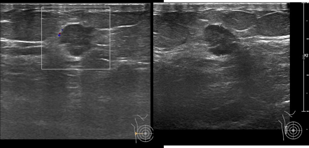
- A lobulated hypoechoic lesion is seen at Rt9H, 5 cm from nipple
- It measures about 9x9x11 mm
- It is taller than its width
- No posterior shadowing seen
- No penetrating vessels
- No abnormal axillary node
Progress of patient:
- A tru-cut biopsy of right breast consistent with invasive carcinoma
- Right mastectomy done
- HPE: invasive carcinoma with no special type, Grade 2
- 12 lymph nodes with no evidence of malignancy
- TNM stage (pTNM) pT1bN0
- ER/Pr +ve and HER2 –ve
- CT scan thorax, abdomen and pelvis show no distant metastasis
Diagnosis: Invasive carcinoma of no special type
Discussion:
- Studies showed DBT has the advantages over FFDM including increased cancer detection
- One of the study reported cancer detection rate in 6 per 1000 DBT compared with 5.1 per 1000 for FFDM alone.
- Screening recall rates were 8% for DBT and 10.4% for FFDM alone
Recent Comments