
Case contribution: Dr Radhiana Hassan
Clinical:
- A 61 years old male with underlying scleroderma under Rheumatologist follow-up
- Presented with progressive swelling over right thumb for 4 years
- Increase in size
- No limitation of movement but patient had intermittent pain
- No clumsiness of hand
- Clinical examination shows a multilobulated swelling over pulp of thumb 2×2 cm, swelling thetered to skin but non-tender.
- Patient is able to flex IPJ and MCPJ. Distal perfusion is good.
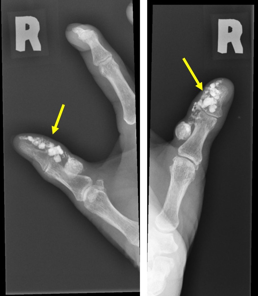
Plain radiograph findings:
- Soft tissue thickening at pulp of right thumb
- Multiple foci of dense calcifications are seen within this thickened soft tissue
- No obvious erosion or periosteal reaction of adjacent phalanges.
- Visualized joints are also normal.
Intra-operative findings:
- One partially liquified lesion over radial side of tip of right thumb excised to its base
- Another hard whitish deposits over ulnar side was also removed
HPE findings:
- Macroscopy: specimen labelled as tumour calcinosis consists of a piece of whitish piece of tissue.
- Microscopy: sections show a cystic lesion lined by a keratinized stratified squamous epithelium. The cavity of the cyst is filled with lamellated keratin flakes. Negative for malignancy.
- Interpretation: Epidermal cyst
Diagnosis: Epidermal inclusion cyst with intralesional calcifications.
Discussion:
- Epidermal inclusion cysts are the most common cutaneous cysts.
- Synonyms include: epidermoid cyst, epidermal cyst, infundibular cyst, inclusion cyst and keratin cyst
- Epidermoids are slow-growing benign cysts bounded by a wall of stratified squamous epithelium
- Epidermoid cyst can occur anywhere in the body bur commonly seen in the scalp, face, neck, trunk and back.
- Less than 10% occur in the extremities
- The size ranges from a few mm to a few cm
- Lesion may remain stable or progressively enlarge over time. No reliable predictive feature to tell if an epidermal inclusion cyst tend to be larger, erythematous or more noticeable to patient
- They do not originate from sebaceous gland, thus are not sebaceous cyst
- Majority are sporadic and not contagious, it can also resolve on their own
- Imaging findings reflects the content of the lesion – debris, water, cholesterol and keratin
- Typically unruptured epidermoid cysts are well-defined round or oval-shaped lesion of high signal intensity on T2WI. However variable signal intensity also reported depending on presence of keratin debri or calcification.
- Malignant degeneration of epidermal cyst is uncommon. Squamous cell carcinoma is seen in 2.2% of cases